 Movie
Movie Controller


+ Open data

- Basic information
| Entry | Database: PDB / ID: 3iys | ||||||
|---|---|---|---|---|---|---|---|
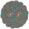
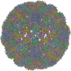
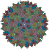
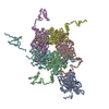
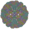
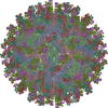
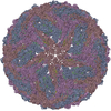
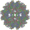
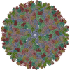
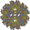
| Title | Homology model of avian polyomavirus asymmetric unit | ||||||
 Components Components | Major capsid protein VP1 | ||||||
 Keywords Keywords | VIRUS / avian / polyomavirus / APV / icosahedral virus | ||||||
| Function / homology |  Function and homology information Function and homology informationT=7 icosahedral viral capsid / endocytosis involved in viral entry into host cell / virion attachment to host cell / host cell nucleus / structural molecule activity Similarity search - Function | ||||||
| Biological species |  Budgerigar fledgling disease polyomavirus Budgerigar fledgling disease polyomavirus | ||||||
| Method | ELECTRON MICROSCOPY / single particle reconstruction / cryo EM / Resolution: 11.3 Å | ||||||
 Authors Authors | Shen, P.S. / Enderlein, D. / Nelson, C.D.S. / Carter, W.S. / Kawano, M. / Xing, L. / Swenson, R.D. / Olson, N.H. / Baker, T.S. / Cheng, R.H. ...Shen, P.S. / Enderlein, D. / Nelson, C.D.S. / Carter, W.S. / Kawano, M. / Xing, L. / Swenson, R.D. / Olson, N.H. / Baker, T.S. / Cheng, R.H. / Atwood, W.J. / Johne, R. / Belnap, D.M. | ||||||
 Citation Citation | Journal: Virology / Year: 2011 Title: The structure of avian polyomavirus reveals variably sized capsids, non-conserved inter-capsomere interactions, and a possible location of the minor capsid protein VP4. Authors: Peter S Shen / Dirk Enderlein / Christian D S Nelson / Weston S Carter / Masaaki Kawano / Li Xing / Robert D Swenson / Norman H Olson / Timothy S Baker / R Holland Cheng / Walter J Atwood / ...Authors: Peter S Shen / Dirk Enderlein / Christian D S Nelson / Weston S Carter / Masaaki Kawano / Li Xing / Robert D Swenson / Norman H Olson / Timothy S Baker / R Holland Cheng / Walter J Atwood / Reimar Johne / David M Belnap /  Abstract: Avian polyomavirus (APV) causes a fatal, multi-organ disease among several bird species. Using cryogenic electron microscopy and other biochemical techniques, we investigated the structure of APV and ...Avian polyomavirus (APV) causes a fatal, multi-organ disease among several bird species. Using cryogenic electron microscopy and other biochemical techniques, we investigated the structure of APV and compared it to that of mammalian polyomaviruses, particularly JC polyomavirus and simian virus 40. The structure of the pentameric major capsid protein (VP1) is mostly conserved; however, APV VP1 has a unique, truncated C-terminus that eliminates an intercapsomere-connecting β-hairpin observed in other polyomaviruses. We postulate that the terminal β-hairpin locks other polyomavirus capsids in a stable conformation and that absence of the hairpin leads to the observed capsid size variation in APV. Plug-like density features were observed at the base of the VP1 pentamers, consistent with the known location of minor capsid proteins VP2 and VP3. However, the plug density is more prominent in APV and may include VP4, a minor capsid protein unique to bird polyomaviruses. | ||||||
| History |
|
- Structure visualization
| Movie |
Movie viewer |
|---|---|
| Structure viewer | Molecule: MolmilJmol/JSmol |
- Downloads & links
-Download
| PDBx/mmCIF format | 3iys.cif.gz | 65 KB | Display | PDBx/mmCIF format |
|---|---|---|---|---|
| PDB format | pdb3iys.ent.gz | 41 KB | Display | PDB format |
| PDBx/mmJSON format | 3iys.json.gz | Tree view | PDBx/mmJSON format | |
| Others |  Other downloads Other downloads |
-Validation report
| Arichive directory | https://data.pdbj.org/pub/pdb/validation_reports/iy/3iysftp://data.pdbj.org/pub/pdb/validation_reports/iy/3iys | HTTPS FTP |
|---|
-Related structure data
| Related structure data | 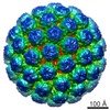 5180MC 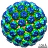 5181C 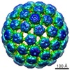 5182C 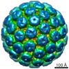 5183C 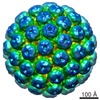 5184C 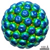 5187C M: map data used to model this data C: citing same article ( |
|---|---|
| Similar structure data |

-Links
 PDBj
PDBj


- Assembly
| Deposited unit | 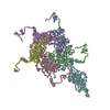
|
|---|---|
| 1 | x 60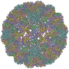
|
| 2 |
|
| 3 | x 5
|
| 4 | x 6
|
| 5 | 
|
| Symmetry | Point symmetry: (Schoenflies symbol: I (icosahedral)) |
-Components
| #1: Protein | Mass: 37449.434 Da / Num. of mol.: 6 / Source method: isolated from a natural source / Source: (natural) Budgerigar fledgling disease polyomavirus / References: UniProt: P13891 |
|---|
-Experimental details
-Experiment
| Experiment | Method: ELECTRON MICROSCOPY |
|---|---|
| EM experiment | Aggregation state: PARTICLE / 3D reconstruction method: single particle reconstruction |
- Sample preparation
| Component | Name: avian polyomavirus / Type: VIRUS / Details: Cryo-EM single particle reconstruction |
|---|---|
| Details of virus | Host category: VERTEBRATES / Isolate: SPECIES / Type: VIRION |
| Natural host | Organism: Aves |
| Buffer solution | Name: GP buffer with 250 mM L-arginine / pH: 10.7 / Details: GP buffer with 250 mM L-arginine |
| Specimen | Embedding applied: NO / Shadowing applied: NO / Staining applied: NO / Vitrification applied: YES |
| Specimen support | Details: avian polyomavirus in holey carbon grid |
| Vitrification | Instrument: FEI VITROBOT MARK I / Cryogen name: ETHANE / Humidity: 100 % / Chamber temperature: 277 K / Details: APV / Method: 3 second blot |
- Electron microscopy imaging
| Experimental equipment |  Model: Tecnai F30 / Image courtesy: FEI Company |
|---|---|
| Microscopy | Model: FEI TECNAI F30 / Date: Apr 23, 2007 |
| Electron gun | Electron source:  FIELD EMISSION GUN / Accelerating voltage: 200 kV / Illumination mode: FLOOD BEAM FIELD EMISSION GUN / Accelerating voltage: 200 kV / Illumination mode: FLOOD BEAM |
| Electron lens | Mode: BRIGHT FIELD / Nominal magnification: 39000 X / Calibrated magnification: 39000 X / Nominal defocus max: 4900 nm / Nominal defocus min: 500 nm Astigmatism: astigmatism was corrected at 59,000 times magnification Camera length: 0 mm |
| Specimen holder | Specimen holder model: GATAN LIQUID NITROGEN Specimen holder type: Side entry liquid nitrogen-cooled cryo specimen holder Temperature: 90 K / Temperature (max): 95 K / Temperature (min): 88 K / Tilt angle max: 0 ° / Tilt angle min: 0 ° |
| Image recording | Film or detector model: KODAK SO-163 FILM |
- Processing
| EM software |
| ||||||||||||
|---|---|---|---|---|---|---|---|---|---|---|---|---|---|
| CTF correction | Details: full CTF correction (FSC cutoff 0.3) | ||||||||||||
| Symmetry | Point symmetry: I (icosahedral) | ||||||||||||
| 3D reconstruction | Method: Fourier Bessel / Resolution: 11.3 Å / Num. of particles: 5338 / Nominal pixel size: 1.63 Å / Actual pixel size: 1.57 Å Magnification calibration: against simian virus 40 atomic coordinates Symmetry type: POINT | ||||||||||||
| Atomic model building | Protocol: RIGID BODY FIT / Space: REAL / Target criteria: cross-correlation coefficient / Details: METHOD--rigid body REFINEMENT PROTOCOL--rigid body | ||||||||||||
| Atomic model building | PDB-ID: 1SIE Accession code: 1SIE / Source name: PDB / Type: experimental model | ||||||||||||
| Refinement step | Cycle: LAST
|
