 Movie
Movie Controller

[English] 日本語
 Yorodumi
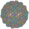
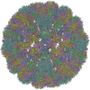
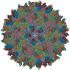
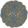
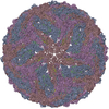
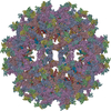
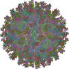
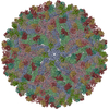
- PDB-7bu8: Cryo-EM structure of zika virus complexed with Fab SIgN-3C at pH 6.5 -
+ Open data

- Basic information
| Entry | Database: PDB / ID: 7bu8 | |||||||||||||||||||||||||||
|---|---|---|---|---|---|---|---|---|---|---|---|---|---|---|---|---|---|---|---|---|---|---|---|---|---|---|---|---|
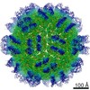
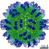
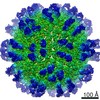
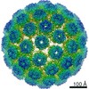
| Title | Cryo-EM structure of zika virus complexed with Fab SIgN-3C at pH 6.5 | |||||||||||||||||||||||||||
 Components Components |
| |||||||||||||||||||||||||||
 Keywords Keywords | VIRUS / Zika virus / antibody / neutralization | |||||||||||||||||||||||||||
| Function / homology |  Function and homology information Function and homology informationflavivirin / symbiont-mediated suppression of host JAK-STAT cascade via inhibition of host TYK2 activity / symbiont-mediated suppression of host JAK-STAT cascade via inhibition of STAT2 activity / symbiont-mediated suppression of host JAK-STAT cascade via inhibition of STAT1 activity / negative regulation of innate immune response / viral capsid / nucleoside-triphosphate phosphatase / double-stranded RNA binding / 4 iron, 4 sulfur cluster binding / clathrin-dependent endocytosis of virus by host cell ...flavivirin / symbiont-mediated suppression of host JAK-STAT cascade via inhibition of host TYK2 activity / symbiont-mediated suppression of host JAK-STAT cascade via inhibition of STAT2 activity / symbiont-mediated suppression of host JAK-STAT cascade via inhibition of STAT1 activity / negative regulation of innate immune response / viral capsid / nucleoside-triphosphate phosphatase / double-stranded RNA binding / 4 iron, 4 sulfur cluster binding / clathrin-dependent endocytosis of virus by host cell / mRNA (guanine-N7)-methyltransferase / methyltransferase cap1 / molecular adaptor activity / methyltransferase cap1 activity / mRNA 5'-cap (guanine-N7-)-methyltransferase activity / RNA helicase activity / protein dimerization activity / host cell perinuclear region of cytoplasm / host cell endoplasmic reticulum membrane / RNA helicase / symbiont-mediated suppression of host type I interferon-mediated signaling pathway / serine-type endopeptidase activity / symbiont-mediated activation of host autophagy / RNA-directed RNA polymerase / viral RNA genome replication / RNA-directed RNA polymerase activity / fusion of virus membrane with host endosome membrane / viral envelope / centrosome / lipid binding / virion attachment to host cell / GTP binding / host cell nucleus / virion membrane / structural molecule activity / ATP hydrolysis activity / proteolysis / extracellular region / ATP binding / metal ion binding Similarity search - Function | |||||||||||||||||||||||||||
| Biological species |  Homo sapiens (human) Homo sapiens (human) Zika virus ZIKV/H. sapiens/FrenchPolynesia/10087PF/2013 Zika virus ZIKV/H. sapiens/FrenchPolynesia/10087PF/2013 | |||||||||||||||||||||||||||
| Method | ELECTRON MICROSCOPY / single particle reconstruction / cryo EM / Resolution: 3.8 Å | |||||||||||||||||||||||||||
 Authors Authors | Zhang, S. / Chew, S.V. / Lim, X.N. / Ng, T.S. / Kostyuchenko, V.A. / Lok, S.M. | |||||||||||||||||||||||||||
| Funding support |  Singapore, 2items Singapore, 2items
| |||||||||||||||||||||||||||
 Citation Citation | Journal: Cell Rep / Year: 2020 Title: A Human Antibody Neutralizes Different Flaviviruses by Using Different Mechanisms. Authors: Shuijun Zhang / Thomas Loy / Thiam-Seng Ng / Xin-Ni Lim / Shyn-Yun Valerie Chew / Ter Yong Tan / Meihui Xu / Victor A Kostyuchenko / Farhana Tukijan / Jian Shi / Katja Fink / Shee-Mei Lok / Abstract: Human antibody SIgN-3C neutralizes dengue virus (DENV) and Zika virus (ZIKV) differently. DENV:SIgN-3C Fab and ZIKV:SIgN-3C Fab cryoelectron microscopy (cryo-EM) complex structures show Fabs ...Human antibody SIgN-3C neutralizes dengue virus (DENV) and Zika virus (ZIKV) differently. DENV:SIgN-3C Fab and ZIKV:SIgN-3C Fab cryoelectron microscopy (cryo-EM) complex structures show Fabs crosslink E protein dimers at extracellular pH 8.0 condition and also when further incubated at acidic endosomal conditions (pH 8.0-6.5). We observe Fab binding to DENV (pH 8.0-5.0) prevents virus fusion, and the number of bound Fabs increase (from 120 to 180). For ZIKV, although there are already 180 copies of Fab at pH 8.0, virus structural changes at pH 5.0 are not inhibited. The immunoglobulin G (IgG):DENV structure at pH 8.0 shows both Fab arms bind to epitopes around the 2-fold vertex. On ZIKV, an additional Fab around the 5-fold vertex at pH 8.0 suggests one IgG arm would engage with an epitope, although the other may bind to other viruses, causing aggregation. For DENV2 at pH 5.0, a similar scenario would occur, suggesting DENV2:IgG complex would aggregate in the endosome. Hence, a single antibody employs different neutralization mechanisms against different flaviviruses. | |||||||||||||||||||||||||||
| History |
|
- Structure visualization
| Movie |
Movie viewer |
|---|---|
| Structure viewer | Molecule: MolmilJmol/JSmol |
- Downloads & links
-Download
| PDBx/mmCIF format | 7bu8.cif.gz | 419.1 KB | Display | PDBx/mmCIF format |
|---|---|---|---|---|
| PDB format | pdb7bu8.ent.gz | 341.3 KB | Display | PDB format |
| PDBx/mmJSON format | 7bu8.json.gz | Tree view | PDBx/mmJSON format | |
| Others |  Other downloads Other downloads |
-Validation report
| Arichive directory | https://data.pdbj.org/pub/pdb/validation_reports/bu/7bu8ftp://data.pdbj.org/pub/pdb/validation_reports/bu/7bu8 | HTTPS FTP |
|---|
-Related structure data
| Related structure data | 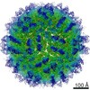 30192MC  7buaC  7bubC  7budC  7bueC  7bufC M: map data used to model this data C: citing same article ( |
|---|---|
| Similar structure data |


-Links
 PDBj
PDBj


- Assembly
| Deposited unit | 
|
|---|---|
| 1 | x 60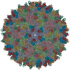
|
| 2 |
|
| 3 | x 5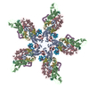
|
| 4 | x 6
|
| 5 | 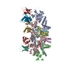
|
| Symmetry | Point symmetry: (Schoenflies symbol: I (icosahedral)) |
-Components
| #1: Protein | Mass: 54444.051 Da / Num. of mol.: 3 / Source method: isolated from a natural source Source: (natural) Zika virus ZIKV/H. sapiens/FrenchPolynesia/10087PF/2013Strain: isolate ZIKV/Human/French Polynesia/10087PF/2013 References: UniProt: A0A024B7W1, flavivirin, nucleoside-triphosphate phosphatase, RNA helicase, mRNA (guanine-N7)-methyltransferase, methyltransferase cap1, RNA-directed RNA polymerase #2: Protein | Mass: 8496.883 Da / Num. of mol.: 3 / Source method: isolated from a natural source Source: (natural) Zika virus ZIKV/H. sapiens/FrenchPolynesia/10087PF/2013Strain: ZIKV/H. sapiens/FrenchPolynesia/10087PF/2013 / References: UniProt: A0A024B7W1*PLUS #3: Antibody | Mass: 14448.958 Da / Num. of mol.: 3 Source method: isolated from a genetically manipulated source Source: (gene. exp.) Homo sapiens (human) / Cell line (production host): HEK293-6E / Production host: Homo sapiens (human)#4: Antibody | Mass: 11832.145 Da / Num. of mol.: 3 Source method: isolated from a genetically manipulated source Source: (gene. exp.) Homo sapiens (human) / Cell line (production host): HEK293-6E / Production host: Homo sapiens (human)#5: Sugar |   Type: D-saccharide, beta linking / Mass: 221.208 Da / Num. of mol.: 3 Type: D-saccharide, beta linking / Mass: 221.208 Da / Num. of mol.: 3Source method: isolated from a genetically manipulated source Formula: C8H15NO6 / Feature type: SUBJECT OF INVESTIGATION Has ligand of interest | Y | Has protein modification | Y | |
|---|
-Experimental details
-Experiment
| Experiment | Method: ELECTRON MICROSCOPY |
|---|---|
| EM experiment | Aggregation state: PARTICLE / 3D reconstruction method: single particle reconstruction |
- Sample preparation
| Component |
| ||||||||||||||||||||||||
|---|---|---|---|---|---|---|---|---|---|---|---|---|---|---|---|---|---|---|---|---|---|---|---|---|---|
| Source (natural) |
| ||||||||||||||||||||||||
| Source (recombinant) | Organism: Homo sapiens (human) / Cell: HEK293-6E | ||||||||||||||||||||||||
| Buffer solution | pH: 6.5 | ||||||||||||||||||||||||
| Specimen | Embedding applied: NO / Shadowing applied: NO / Staining applied: NO / Vitrification applied: YES | ||||||||||||||||||||||||
| Vitrification | Cryogen name: ETHANE |
- Electron microscopy imaging
| Experimental equipment |  Model: Titan Krios / Image courtesy: FEI Company |
|---|---|
| Microscopy | Model: FEI TITAN KRIOS |
| Electron gun | Electron source:  FIELD EMISSION GUN / Accelerating voltage: 300 kV / Illumination mode: FLOOD BEAM FIELD EMISSION GUN / Accelerating voltage: 300 kV / Illumination mode: FLOOD BEAM |
| Electron lens | Mode: BRIGHT FIELD |
| Image recording | Electron dose: 30 e/Å2 / Film or detector model: FEI FALCON II (4k x 4k) |
- Processing
| Software | Name: PHENIX / Version: 1.12_2829: / Classification: refinement | ||||||||||||||||||||||||
|---|---|---|---|---|---|---|---|---|---|---|---|---|---|---|---|---|---|---|---|---|---|---|---|---|---|
| EM software | Name: PHENIX / Category: model refinement | ||||||||||||||||||||||||
| CTF correction | Type: PHASE FLIPPING AND AMPLITUDE CORRECTION | ||||||||||||||||||||||||
| 3D reconstruction | Resolution: 3.8 Å / Resolution method: FSC 0.143 CUT-OFF / Num. of particles: 16300 / Symmetry type: POINT | ||||||||||||||||||||||||
| Refine LS restraints |
|
