 Movie
Movie Controller


+ Open data

- Basic information
| Entry | Database: EMDB / ID: EMD-11293 | ||||||||||||
|---|---|---|---|---|---|---|---|---|---|---|---|---|---|
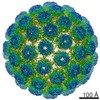
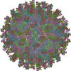
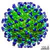
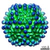
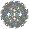
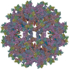
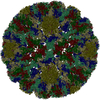
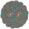
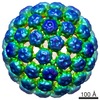
| Title | CryoEM Structure of Merkel Cell Polyomavirus | ||||||||||||
 Map data Map data | |||||||||||||
 Sample Sample |
| ||||||||||||
 Keywords Keywords | polyomavirus / capsid / Merkel cell carcinoma / jelly roll fold / VIRUS LIKE PARTICLE | ||||||||||||
| Function / homology |  Function and homology information Function and homology informationT=7 icosahedral viral capsid / endocytosis involved in viral entry into host cell / virion attachment to host cell / host cell nucleus / structural molecule activity / metal ion binding Similarity search - Function | ||||||||||||
| Biological species |  Merkel cell polyomavirus Merkel cell polyomavirus | ||||||||||||
| Method | single particle reconstruction / cryo EM / Resolution: 3.4 Å | ||||||||||||
 Authors Authors | Bayer NJ / Januliene D | ||||||||||||
| Funding support |  Germany, 3 items Germany, 3 items
| ||||||||||||
 Citation Citation | Journal: J Virol / Year: 2020 Title: Structure of Merkel Cell Polyomavirus Capsid and Interaction with Its Glycosaminoglycan Attachment Receptor. Authors: Niklas J Bayer / Dovile Januliene / Georg Zocher / Thilo Stehle / Arne Moeller / Bärbel S Blaum /  Abstract: Merkel cell polyomavirus (MCPyV) is a human double-stranded DNA tumor virus. MCPyV cell entry is unique among members of the polyomavirus family as it requires the engagement of two types of glycans, ...Merkel cell polyomavirus (MCPyV) is a human double-stranded DNA tumor virus. MCPyV cell entry is unique among members of the polyomavirus family as it requires the engagement of two types of glycans, sialylated oligosaccharides and sulfated glycosaminoglycans (GAGs). Here, we present crystallographic and cryo-electron microscopic structures of the icosahedral MCPyV capsid and analysis of its glycan interactions via nuclear magnetic resonance (NMR) spectroscopy. While sialic acid binding is specific for α2-3-linked sialic acid and mediated by the exposed apical loops of the major capsid protein VP1, a broad range of GAG oligosaccharides bind to recessed regions between VP1 capsomers. Individual VP1 capsomers are tethered to one another by an extensive disulfide network that differs in architecture from previously described interactions for other PyVs. An unusual C-terminal extension in MCPyV VP1 projects from the recessed capsid regions. Mutagenesis experiments show that this extension is dispensable for receptor interactions. The MCPyV genome was found to be clonally integrated in 80% of cases of Merkel cell carcinoma (MCC), a rare but aggressive form of human skin cancer, strongly suggesting that this virus is tumorigenic. In the metastasizing state, the course of the disease is often fatal, especially in immunocompromised individuals, as reflected by the high mortality rate of 33 to 46% and the low 5-year survival rate (<45%). The high seroprevalence of about 60% makes MCPyV a serious health care burden and illustrates the need for targeted treatments. In this study, we present the first high-resolution structural data for this human tumor virus and demonstrate that the full capsid is required for the essential interaction with its GAG receptor(s). Together, these data can be used as a basis for future strategies in drug development. | ||||||||||||
| History |
|
- Structure visualization
| Movie |
Movie viewer |
|---|---|
| Structure viewer | EM map: SurfViewMolmilJmol/JSmol |
| Supplemental images |

- Downloads & links
-EMDB archive
| Map data | emd_11293.map.gz | 523.1 MB | EMDB map data format | |
|---|---|---|---|---|
| Header (meta data) | emd-11293-v30.xmlemd-11293.xml | 21.7 KB 21.7 KB | Display Display | EMDB header |
| FSC (resolution estimation) | emd_11293_fsc.xml | 24.1 KB | Display | FSC data file |
| Images | 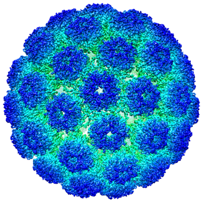 emd_11293.png emd_11293.png | 295.6 KB | ||
| Filedesc metadata | emd-11293.cif.gz | 6.1 KB | ||
| Others | emd_11293_additional.map.gzemd_11293_half_map_1.map.gzemd_11293_half_map_2.map.gz | 518.7 MB 212 MB 212 MB | ||
| Archive directory |  http://ftp.pdbj.org/pub/emdb/structures/EMD-11293ftp://ftp.pdbj.org/pub/emdb/structures/EMD-11293 http://ftp.pdbj.org/pub/emdb/structures/EMD-11293ftp://ftp.pdbj.org/pub/emdb/structures/EMD-11293 | HTTPS FTP |
-Related structure data
| Related structure data |  6zmlMC  6zlzC M: atomic model generated by this map C: citing same article ( |
|---|---|
| Similar structure data |
-Links
| EMDB pages | EMDB (EBI/PDBe) / EMDataResource |
|---|---|
| Related items in Molecule of the Month |


-Map
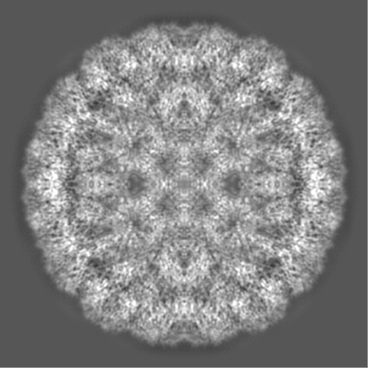
| File | Download / File: emd_11293.map.gz / Format: CCP4 / Size: 561.5 MB / Type: IMAGE STORED AS FLOATING POINT NUMBER (4 BYTES) | ||||||||||||||||||||||||||||||||||||||||||||||||||||||||||||
|---|---|---|---|---|---|---|---|---|---|---|---|---|---|---|---|---|---|---|---|---|---|---|---|---|---|---|---|---|---|---|---|---|---|---|---|---|---|---|---|---|---|---|---|---|---|---|---|---|---|---|---|---|---|---|---|---|---|---|---|---|---|
| Projections & slices | Image control
Images are generated by Spider. | ||||||||||||||||||||||||||||||||||||||||||||||||||||||||||||
| Voxel size | X=Y=Z: 1.053 Å | ||||||||||||||||||||||||||||||||||||||||||||||||||||||||||||
| Density |
| ||||||||||||||||||||||||||||||||||||||||||||||||||||||||||||
| Symmetry | Space group: 1 | ||||||||||||||||||||||||||||||||||||||||||||||||||||||||||||
| Details | EMDB XML:
CCP4 map header:
| ||||||||||||||||||||||||||||||||||||||||||||||||||||||||||||
 Z (Sec.)
 Y (Row.)
 X (Col.)


-Supplemental data
-Additional map: Merkel Cell polyomavirus (unsharpened map).
| File | emd_11293_additional.map | ||||||||||||
|---|---|---|---|---|---|---|---|---|---|---|---|---|---|
| Annotation | Merkel Cell polyomavirus (unsharpened map). | ||||||||||||
| Projections & Slices |
| ||||||||||||
| Density Histograms |


-Half map: #1
| File | emd_11293_half_map_1.map | ||||||||||||
|---|---|---|---|---|---|---|---|---|---|---|---|---|---|
| Projections & Slices |
| ||||||||||||
| Density Histograms |


-Half map: #2
| File | emd_11293_half_map_2.map | ||||||||||||
|---|---|---|---|---|---|---|---|---|---|---|---|---|---|
| Projections & Slices |
| ||||||||||||
| Density Histograms |


- Sample components
-Entire : Merkel cell polyomavirus
| Entire | Name: Merkel cell polyomavirus |
|---|---|
| Components |
|
-Supramolecule #1: Merkel cell polyomavirus
| Supramolecule | Name: Merkel cell polyomavirus / type: virus / ID: 1 / Parent: 0 / Macromolecule list: all / NCBI-ID: 493803 / Sci species name: Merkel cell polyomavirus / Virus type: VIRUS-LIKE PARTICLE / Virus isolate: OTHER / Virus enveloped: No / Virus empty: Yes |
|---|---|
| Host (natural) | Organism:  Homo sapiens (human) Homo sapiens (human) |
| Virus shell | Shell ID: 1 / Diameter: 508.0 Å / T number (triangulation number): 7 |
-Macromolecule #1: Capsid protein VP1
| Macromolecule | Name: Capsid protein VP1 / type: protein_or_peptide / ID: 1 / Number of copies: 6 / Enantiomer: LEVO |
|---|---|
| Source (natural) | Organism: Merkel cell polyomavirus |
| Molecular weight | Theoretical: 46.611031 KDa |
| Recombinant expression | Organism: Homo sapiens (human) |
| Sequence | String: MAPKRKASST CKTPKRQCIP KPGCCPNVAS VPKLLVKGGV EVLSVVTGED SITQIELYLN PRMGVNSPDL PTTSNWYTYT YDLQPKGSS PDQPIKENLP AYSVARVSLP MLNEDITCDT LQMWEAISVK TEVVGISSLI NVHYWDMKRV HDYGAGIPVS G VNYHMFAI ...String: MAPKRKASST CKTPKRQCIP KPGCCPNVAS VPKLLVKGGV EVLSVVTGED SITQIELYLN PRMGVNSPDL PTTSNWYTYT YDLQPKGSS PDQPIKENLP AYSVARVSLP MLNEDITCDT LQMWEAISVK TEVVGISSLI NVHYWDMKRV HDYGAGIPVS G VNYHMFAI GGEPLDLQGL VLDYQTQYPK TTNGGPITIE TVLGRKMTPK NQGLDPQAKA KLDKDGNYPI EVWCPDPSKN EN SRYYGSI QTGSQTPTVL QFSNTLTTVL LDENGVGPLC KGDGLFISCA DIVGFLFKTS GKMALHGLPR YFNVTLRKRW VKN PYPVVN LINSLFSNLM PKVSGQPMEG KDNQVEEVRI YEGSEQLPGD PDIVRFLDKF GQEKTVYPKP SVAPAAVTFQ SNQQ DKGKA PLKGPQKASQ KESQTQEL UniProtKB: VP1 |
-Experimental details
-Structure determination
| Method | cryo EM |
|---|---|
 Processing Processing | single particle reconstruction |
| Aggregation state | particle |
-Sample preparation
| Concentration | 1 mg/mL | ||||||||||||
|---|---|---|---|---|---|---|---|---|---|---|---|---|---|
| Buffer | pH: 6.6 Component:
Details: Solution was sterile filtered | ||||||||||||
| Grid | Model: C-flat-2/2 / Material: COPPER / Pretreatment - Type: GLOW DISCHARGE | ||||||||||||
| Vitrification | Cryogen name: ETHANE / Chamber humidity: 100 % / Chamber temperature: 277 K / Instrument: FEI VITROBOT MARK IV |
- Electron microscopy
| Microscope | FEI TITAN KRIOS |
|---|---|
| Image recording | Film or detector model: FEI FALCON III (4k x 4k) / Detector mode: COUNTING / Number grids imaged: 1 / Number real images: 463 / Average exposure time: 67.0 sec. / Average electron dose: 30.0 e/Å2 |
| Electron beam | Acceleration voltage: 300 kV / Electron source:  FIELD EMISSION GUN FIELD EMISSION GUN |
| Electron optics | Illumination mode: FLOOD BEAM / Imaging mode: BRIGHT FIELD / Nominal defocus max: 1.5 µm / Nominal defocus min: 0.5 µm / Nominal magnification: 75000 |
| Sample stage | Cooling holder cryogen: NITROGEN |
| Experimental equipment |  Model: Titan Krios / Image courtesy: FEI Company |
+Image processing

-Atomic model buiding 1
| Initial model |
| ||||||||||||||
|---|---|---|---|---|---|---|---|---|---|---|---|---|---|---|---|
| Refinement | Space: REAL / Protocol: FLEXIBLE FIT / Overall B value: 105.92 / Target criteria: Correlation coefficient | ||||||||||||||
| Output model | PDB-6zml: |
