 Movie
Movie Controller


+ Open data

- Basic information
| Entry | Database: PDB / ID: 1sid | |||||||||
|---|---|---|---|---|---|---|---|---|---|---|
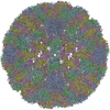
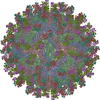
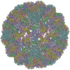
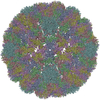
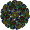
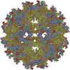
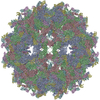
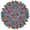
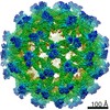
| Title | MURINE POLYOMAVIRUS COMPLEXED WITH 3'SIALYL LACTOSE | |||||||||
 Components Components | POLYOMAVIRUS COAT PROTEIN VP1 | |||||||||
 Keywords Keywords | VIRUS / COAT PROTEIN / Icosahedral virus | |||||||||
| Function / homology |  Function and homology information Function and homology informationcaveolin-mediated endocytosis of virus by host cell / T=7 icosahedral viral capsid / virion attachment to host cell / host cell nucleus / structural molecule activity Similarity search - Function | |||||||||
| Biological species |  Mouse polyomavirus Mouse polyomavirus | |||||||||
| Method |  X-RAY DIFFRACTION / Resolution: 3.65 Å X-RAY DIFFRACTION / Resolution: 3.65 Å | |||||||||
 Authors Authors | Stehle, T. / Harrison, S.C. | |||||||||
 Citation Citation | Journal: Structure / Year: 1996 Title: Crystal structures of murine polyomavirus in complex with straight-chain and branched-chain sialyloligosaccharide receptor fragments. Authors: Stehle, T. / Harrison, S.C. #1: Journal: To be PublishedTitle: The Structure of Simian Virus 40 Refined at 3.1 Angstroms Resolution Authors: Stehle, T. / Gamblin, S.J. / Yan, Y. / Harrison, S.C. #2: Journal: To be PublishedTitle: Structure Determination of Simian Virus 40 and Murine Polyomavirus by a Combination of 5-Fold and 30-Fold Electron Density Averaging Authors: Yan, Y. / Stehle, T. / Liddington, R.C. / Zhao, H.C. / Harrison, S.C. #3: Journal: Nature / Year: 1994Title: Structure of Murine Polyomavirus Complexed with an Oligosaccharide Receptor Fragment Authors: Stehle, T. / Yan, Y. / Benjamin, T.L. / Harrison, S.C. | |||||||||
| History |
|
- Structure visualization
| Structure viewer | Molecule: MolmilJmol/JSmol |
|---|
- Downloads & links
-Download
| PDBx/mmCIF format | 1sid.cif.gz | 427.8 KB | Display | PDBx/mmCIF format |
|---|---|---|---|---|
| PDB format | pdb1sid.ent.gz | 354.1 KB | Display | PDB format |
| PDBx/mmJSON format | 1sid.json.gz | Tree view | PDBx/mmJSON format | |
| Others |  Other downloads Other downloads |
-Validation report
| Arichive directory | https://data.pdbj.org/pub/pdb/validation_reports/si/1sidftp://data.pdbj.org/pub/pdb/validation_reports/si/1sid | HTTPS FTP |
|---|
-Related structure data

-Links
 PDBj
PDBj


- Assembly
| Deposited unit | 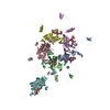
| ||||||||||||||||||
|---|---|---|---|---|---|---|---|---|---|---|---|---|---|---|---|---|---|---|---|
| 1 | x 60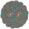
| ||||||||||||||||||
| 2 |
| ||||||||||||||||||
| 3 | x 5
| ||||||||||||||||||
| 4 | x 6
| ||||||||||||||||||
| 5 | 
| ||||||||||||||||||
| 6 | x 5
| ||||||||||||||||||
| Unit cell |
| ||||||||||||||||||
| Symmetry | Point symmetry: (Hermann–Mauguin notation: 532 / Schoenflies symbol: I (icosahedral)) | ||||||||||||||||||
| Noncrystallographic symmetry (NCS) | NCS oper:
| ||||||||||||||||||
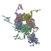
| Details | SPACE GROUP I23, A=570 ANG. THERE ARE TWO VIRIONS PER UNIT CELL AND 30 VP1 MONOMERS IN THE CRYSTALLOGRAPHIC ASYMMETRIC UNIT. IN THIS STRUCTURE THERE ARE INVADING ARMS OF SYMMETRY-RELATED NEIGHBORING MONOMERS THAT INVADE THE REFERENCE PENTAMER. IN REFINEMENT, THE AUTHORS USED RESIDUES 337 TO THE C-TERMINUS FROM THE SYMMETRY-RELATED MONOMERS. IN THIS ENTRY, RESIDUES 337 TO THE C-TERMINUS ARE PRESENTED AS THE ACTUAL ARM OF THE MONOMER (THAT INVADES ANOTHER PENTAMER). BECAUSE THE REFINEMENT DID NOT USE A BOND BETWEEN RESIDUES 336 AND 337 (BECAUSE THEY BELONGED TO DIFFERENT CHAINS), THE GEOMETRY OF THAT BOND IS NOT ACCURATE. A STRICT PENTAMER SURROUNDED BY FIVE LOCAL PENTAMERS CONSTITUTES THE CRYSTALLOGRAPHIC ASYMMETRIC UNIT. APPLICATION OF THE FIRST FIVE MATRICES TO THE COORDINATE SET GENERATES THAT CRYSTALLOGRAPHIC ASYMMETRIC UNIT. |
-Components
| #1: Protein | Mass: 42405.109 Da / Num. of mol.: 6 / Source method: isolated from a natural source Source: (natural) Mouse polyomavirus (strain p16 small-plaque)Genus: Polyomavirus / Species: Murine polyomavirus / Strain: SMALL-PLAQUE P16 / References: UniProt: P49302 #2: Polysaccharide | N-acetyl-alpha-neuraminic acid-(2-3)-beta-D-galactopyranose-(1-4)-beta-D-glucopyranose Source method: isolated from a genetically manipulated source Has protein modification | Y | |
|---|
-Experimental details
-Experiment
| Experiment | Method: X-RAY DIFFRACTION |
|---|
- Sample preparation
| Crystal grow | *PLUS pH: 7.5 / Method: vapor diffusion, hanging drop | ||||||||||||||||||||||||||||||||||||||||
|---|---|---|---|---|---|---|---|---|---|---|---|---|---|---|---|---|---|---|---|---|---|---|---|---|---|---|---|---|---|---|---|---|---|---|---|---|---|---|---|---|---|
| Components of the solutions | *PLUS
|
-Data collection
| Radiation | Scattering type: x-ray |
|---|---|
| Radiation wavelength | Relative weight: 1 |
| Reflection | Resolution: 3.65→12 Å / Num. obs: 242373 / % possible obs: 73 % / Observed criterion σ(I): 0 |
- Processing
| Software |
| ||||||||||||||||
|---|---|---|---|---|---|---|---|---|---|---|---|---|---|---|---|---|---|
| Refinement | Resolution: 3.65→12 Å / σ(F): 0
| ||||||||||||||||
| Refinement step | Cycle: LAST / Resolution: 3.65→12 Å
|
