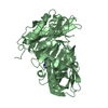
Entry Database : PDB / ID : 3iw4Title Crystal structure of PKC alpha in complex with NVP-AEB071 Protein kinase C alpha type Keywords / / / / / / / / / / Function / homology Function Domain/homology Component
/ / / / / / / / / / / / / / / / / / / / / / / / / / / / / / / / / / / / / / / / / / / / / / / / / / / / / / / / / / / / / / / / / / / / / / / / / / / / / / / / / / / / / / / / / / / / / / / / / / / / / / / / / / / / / / / / / / / / / / / / / / / / / / / / / / / / / / / / / / / / Biological species Homo sapiens (human)Method / / / Resolution : 2.8 Å Authors Stark, W. / Rummel, G. / Strauss, A. / Cowan-Jacob, S.W. Journal : J.Med.Chem. / Year : 2009Title : Discovery of 3-(1H-indol-3-yl)-4-[2-(4-methylpiperazin-1-yl)quinazolin-4-yl]pyrrole-2,5-dione (AEB071), a potent and selective inhibitor of protein kinase C isotypesAuthors: Wagner, J. / von Matt, P. / Sedrani, R. / Albert, R. / Cooke, N. / Ehrhardt, C. / Geiser, M. / Rummel, G. / Stark, W. / Strauss, A. / Cowan-Jacob, S.W. / Beerli, C. / Weckbecker, G. / ... Authors : Wagner, J. / von Matt, P. / Sedrani, R. / Albert, R. / Cooke, N. / Ehrhardt, C. / Geiser, M. / Rummel, G. / Stark, W. / Strauss, A. / Cowan-Jacob, S.W. / Beerli, C. / Weckbecker, G. / Evenou, J.P. / Zenke, G. / Cottens, S. History Deposition Sep 2, 2009 Deposition site / Processing site Revision 1.0 Nov 3, 2009 Provider / Type Revision 1.1 Jul 13, 2011 Group / Refinement description / Version format complianceRevision 1.2 Nov 1, 2017 Group / Category / Item Revision 1.3 Nov 10, 2021 Group / Derived calculationsCategory database_2 / struct_conn ... database_2 / struct_conn / struct_ref_seq_dif / struct_site Item _database_2.pdbx_DOI / _database_2.pdbx_database_accession ... _database_2.pdbx_DOI / _database_2.pdbx_database_accession / _struct_conn.pdbx_leaving_atom_flag / _struct_ref_seq_dif.details / _struct_site.pdbx_auth_asym_id / _struct_site.pdbx_auth_comp_id / _struct_site.pdbx_auth_seq_id Revision 1.4 Nov 20, 2024 Group / Structure summaryCategory chem_comp_atom / chem_comp_bond ... chem_comp_atom / chem_comp_bond / pdbx_entry_details / pdbx_modification_feature
Show all Show less
 Movie
Movie Controller


 Open data
 Basic information
 Components
 Keywords
 Function and homology information
 Homo sapiens (human)
 X-RAY DIFFRACTION /
  Authors
 Citation
 Structure visualization
 Downloads & links
 Other downloads
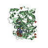


 PDBj
PDBj


 Assembly
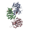


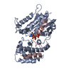
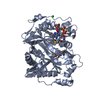
 Spodoptera frugiperda (fall armyworm) / References: UniProt: P17252, protein kinase C
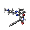
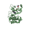
 Mass: 438.481 Da / Num. of mol.: 3 / Source method: obtained synthetically / Formula: C25H22N6O2
 Mass: 18.015 Da / Num. of mol.: 46 / Source method: isolated from a natural source / Formula: H2O
 Sample preparation
 / Beamline: X10SA / Wavelength: 0.9788 Å
 Processing