- PDB-1wmk: Human death-associated kinase DRP-1, mutant S308D d40 -
+
Open data
ID or keywords:
Loading...
-
Basic information
Entry
Database: PDB / ID: 1wmk
Title
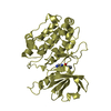
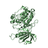
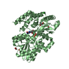
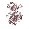
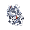
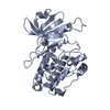
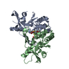
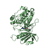
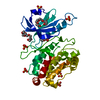
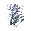
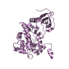
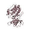
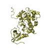
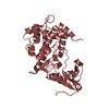
Human death-associated kinase DRP-1, mutant S308D d40
Components
Death-associated protein kinase 2
Keywords
TRANSFERASE / protein kinase / autoinhibitory helix
Function / homology
Function and homology information
positive regulation of eosinophil chemotaxis / autophagosome lumen / regulation of intrinsic apoptotic signaling pathway / neutrophil migration / Caspase activation via Dependence Receptors in the absence of ligand / anoikis / positive regulation of neutrophil chemotaxis / regulation of autophagy / protein autophosphorylation / cytoplasmic vesicle ...positive regulation of eosinophil chemotaxis / autophagosome lumen / regulation of intrinsic apoptotic signaling pathway / neutrophil migration / Caspase activation via Dependence Receptors in the absence of ligand / anoikis / positive regulation of neutrophil chemotaxis / regulation of autophagy / protein autophosphorylation / cytoplasmic vesicle / regulation of apoptotic process / protein phosphorylation / calmodulin binding / non-specific serine/threonine protein kinase / intracellular signal transduction / positive regulation of apoptotic process / protein serine kinase activity / protein serine/threonine kinase activity / apoptotic process / Golgi apparatus / ATP binding / identical protein binding / nucleus / cytoplasm Similarity search - Function
Single helix bin / Single alpha-helices involved in coiled-coils or other helix-helix interfaces / Phosphorylase Kinase; domain 1 / Phosphorylase Kinase; domain 1 / Transferase(Phosphotransferase) domain 1 / Transferase(Phosphotransferase); domain 1 / Serine/threonine-protein kinase, active site / Serine/Threonine protein kinases active-site signature. / Protein kinase domain / Serine/Threonine protein kinases, catalytic domain ...Single helix bin / Single alpha-helices involved in coiled-coils or other helix-helix interfaces / Phosphorylase Kinase; domain 1 / Phosphorylase Kinase; domain 1 / Transferase(Phosphotransferase) domain 1 / Transferase(Phosphotransferase); domain 1 / Serine/threonine-protein kinase, active site / Serine/Threonine protein kinases active-site signature. / Protein kinase domain / Serine/Threonine protein kinases, catalytic domain / Protein kinase, ATP binding site / Protein kinases ATP-binding region signature. / Protein kinase domain profile. / Protein kinase domain / Protein kinase-like domain superfamily / Up-down Bundle / 2-Layer Sandwich / Orthogonal Bundle / Mainly Alpha / Alpha Beta Similarity search - Domain/homology
A: Death-associated protein kinase 2 E: Death-associated protein kinase 2 C: Death-associated protein kinase 2 B: Death-associated protein kinase 2 F: Death-associated protein kinase 2 D: Death-associated protein kinase 2 H: Death-associated protein kinase 2 G: Death-associated protein kinase 2
Resolution: 3.6→20 Å / Cor.coef. Fo:Fc: 0.886 / Cor.coef. Fo:Fc free: 0.873 / SU B: 53.183 / SU ML: 0.732 Isotropic thermal model: GROUP B FACTOR REFINEMENT IN CNS, OVERALL B FACTOR REFINEMENT IN REFMAC5 Cross valid method: THROUGHOUT / σ(F): -3 / ESU R Free: 0.814 / Stereochemistry target values: MAXIMUM LIKELIHOOD Details: HYDROGENS HAVE BEEN ADDED IN THE RIDING POSITIONS. GROUP B FACTOR REFINEMENT IN CNS, OVERALL B FACTOR REFINEMENT IN REFMAC5
Rfactor
Num. reflection
% reflection
Selection details
Rfree
0.29577
1864
5 %
RANDOM
Rwork
0.27633
-
-
-
all
0.2773
37254
-
-
obs
0.2773
37254
99.97 %
-
Solvent computation
Ion probe radii: 0.8 Å / Shrinkage radii: 0.8 Å / VDW probe radii: 1.2 Å / Solvent model: MASK
In the structure databanks used in Yorodumi, some data are registered as the other names, "COVID-19 virus" and "2019-nCoV". Here are the details of the virus and the list of structure data.
Jan 31, 2019. EMDB accession codes are about to change! (news from PDBe EMDB page)
EMDB accession codes are about to change! (news from PDBe EMDB page)
The allocation of 4 digits for EMDB accession codes will soon come to an end. Whilst these codes will remain in use, new EMDB accession codes will include an additional digit and will expand incrementally as the available range of codes is exhausted. The current 4-digit format prefixed with “EMD-” (i.e. EMD-XXXX) will advance to a 5-digit format (i.e. EMD-XXXXX), and so on. It is currently estimated that the 4-digit codes will be depleted around Spring 2019, at which point the 5-digit format will come into force.
The EM Navigator/Yorodumi systems omit the EMD- prefix.
Related info.:Q: What is EMD? / ID/Accession-code notation in Yorodumi/EM Navigator
Yorodumi is a browser for structure data from EMDB, PDB, SASBDB, etc.
This page is also the successor to EM Navigator detail page, and also detail information page/front-end page for Omokage search.
The word "yorodu" (or yorozu) is an old Japanese word meaning "ten thousand". "mi" (miru) is to see.
Related info.:EMDB / PDB / SASBDB / Comparison of 3 databanks / Yorodumi Search / Aug 31, 2016. New EM Navigator & Yorodumi / Yorodumi Papers / Jmol/JSmol / Function and homology information / Changes in new EM Navigator and Yorodumi
 Movie
Movie Controller


 Open data
 Basic information
 Components
 Keywords
 Function and homology information
 Homo sapiens (human)
 X-RAY DIFFRACTION /
  Authors
 Citation
 Structure visualization
 Downloads & links
 Other downloads

 PDBj
PDBj
 Assembly


 Sample preparation
 / Beamline: X13
 Processing