 Movie
Movie Controller

[English] 日本語
 Yorodumi
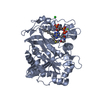
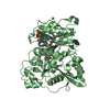
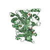
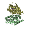
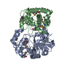
- PDB-3g9k: Crystal structure of Bacillus anthracis transpeptidase enzyme CapD -
+ Open data

- Basic information
| Entry | Database: PDB / ID: 3g9k | ||||||
|---|---|---|---|---|---|---|---|
| Title | Crystal structure of Bacillus anthracis transpeptidase enzyme CapD | ||||||
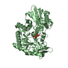
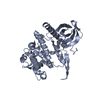
 Components Components | (Capsule biosynthesis protein capD) x 2 | ||||||
 Keywords Keywords | HYDROLASE / CapD protein / Bacillus anthracis / The Great Lakes Regional Center of Excellence / GLRCE / Capsule biogenesis/degradation / Virulence | ||||||
| Function / homology |  Function and homology information Function and homology informationcapsule polysaccharide biosynthetic process / Transferases; Acyltransferases; Aminoacyltransferases / peptidase activity / transferase activity / proteolysis Similarity search - Function | ||||||
| Biological species |  | ||||||
| Method |  X-RAY DIFFRACTION / SYNCHROTRON / MOLECULAR REPLACEMENT / Resolution: 1.79 Å X-RAY DIFFRACTION / SYNCHROTRON / MOLECULAR REPLACEMENT / Resolution: 1.79 Å | ||||||
 Authors Authors | Zhang, R. / Wu, R. / Richter, S. / Anderson, V.J. / Missiakas, D. / Joachimiak, A. | ||||||
 Citation Citation | Journal: J.Biol.Chem. / Year: 2009 Title: Crystal Structure of Bacillus anthracis Transpeptidase Enzyme CapD. Authors: Wu, R. / Richter, S. / Zhang, R.G. / Anderson, V.J. / Missiakas, D. / Joachimiak, A. | ||||||
| History |
|
- Structure visualization
| Structure viewer | Molecule: MolmilJmol/JSmol |
|---|
- Downloads & links
-Download
| PDBx/mmCIF format | 3g9k.cif.gz | 198.8 KB | Display | PDBx/mmCIF format |
|---|---|---|---|---|
| PDB format | pdb3g9k.ent.gz | 156.8 KB | Display | PDB format |
| PDBx/mmJSON format | 3g9k.json.gz | Tree view | PDBx/mmJSON format | |
| Others |  Other downloads Other downloads |
-Validation report
| Arichive directory | https://data.pdbj.org/pub/pdb/validation_reports/g9/3g9kftp://data.pdbj.org/pub/pdb/validation_reports/g9/3g9k | HTTPS FTP |
|---|
-Related structure data
| Related structure data |  3ga9SC S: Starting model for refinement C: citing same article ( |
|---|---|
| Similar structure data |


-Links
 PDBj
PDBj


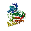
- Assembly
| Deposited unit | 
| ||||||||
|---|---|---|---|---|---|---|---|---|---|
| 1 | 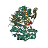
| ||||||||
| 2 | 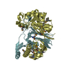
| ||||||||
| Unit cell |
|
-Components
| #1: Protein | Mass: 36223.242 Da / Num. of mol.: 2 / Fragment: UNP residues 29-351 Source method: isolated from a genetically manipulated source Details: Long chain / Source: (gene. exp.) Gene: BAK_B0097, BXB0063, capD, dep, GBAA_pXO2_0063, pXO2-55 Plasmid: pDM68 / Production host: #2: Protein | Mass: 19594.777 Da / Num. of mol.: 2 / Fragment: UNP residues 352-528 Source method: isolated from a genetically manipulated source Details: Short chain / Source: (gene. exp.) #3: Chemical |   Type: L-peptide linking / Mass: 147.129 Da / Num. of mol.: 2 / Source method: obtained synthetically / Formula: C5H9NO4 Type: L-peptide linking / Mass: 147.129 Da / Num. of mol.: 2 / Source method: obtained synthetically / Formula: C5H9NO4#4: Water | ChemComp-HOH / |  Mass: 18.015 Da / Num. of mol.: 555 / Source method: isolated from a natural source / Formula: H2O Mass: 18.015 Da / Num. of mol.: 555 / Source method: isolated from a natural source / Formula: H2OHas protein modification | Y | |
|---|
-Experimental details
-Experiment
| Experiment | Method: X-RAY DIFFRACTION / Number of used crystals: 1 |
|---|
- Sample preparation
| Crystal | Density Matthews: 2.2 Å3/Da / Density % sol: 44.12 % |
|---|---|
| Crystal grow | Temperature: 298 K / Method: vapor diffusion, sitting drop / pH: 7 Details: 15% PEG 3350, 8% PEG 400, 0.05M LiSO4, 0.1M Na-Hepes pH 7.0, VAPOR DIFFUSION, SITTING DROP, temperature 298K |
-Data collection
| Diffraction | Mean temperature: 100 K |
|---|---|
| Diffraction source | Source: SYNCHROTRON / Site: APS  / Beamline: 19-ID / Wavelength: 0.9794 Å / Beamline: 19-ID / Wavelength: 0.9794 Å |
| Detector | Type: ADSC QUANTUM 315 / Detector: CCD / Date: Dec 19, 2006 / Details: mirrors |
| Radiation | Monochromator: Si(111) channel / Protocol: SINGLE WAVELENGTH / Monochromatic (M) / Laue (L): M / Scattering type: x-ray |
| Radiation wavelength | Wavelength: 0.9794 Å / Relative weight: 1 |
| Reflection | Resolution: 1.79→50 Å / Num. all: 88013 / Num. obs: 83551 / % possible obs: 98 % / Redundancy: 4.7 % / Rmerge(I) obs: 0.117 / Net I/σ(I): 16.1 |
| Reflection shell | Resolution: 1.79→1.83 Å / Redundancy: 4 % / Rmerge(I) obs: 0.555 / Mean I/σ(I) obs: 1.73 / % possible all: 82.9 |
- Processing
| Software |
| ||||||||||||||||||||||||||||||||||||||||||||||||||||||||||||||||||||||||||||||||||||||||||||||||||||||||||||||||||||||||||||||||||||||||||||||||||||||||||||||||||||||||||
|---|---|---|---|---|---|---|---|---|---|---|---|---|---|---|---|---|---|---|---|---|---|---|---|---|---|---|---|---|---|---|---|---|---|---|---|---|---|---|---|---|---|---|---|---|---|---|---|---|---|---|---|---|---|---|---|---|---|---|---|---|---|---|---|---|---|---|---|---|---|---|---|---|---|---|---|---|---|---|---|---|---|---|---|---|---|---|---|---|---|---|---|---|---|---|---|---|---|---|---|---|---|---|---|---|---|---|---|---|---|---|---|---|---|---|---|---|---|---|---|---|---|---|---|---|---|---|---|---|---|---|---|---|---|---|---|---|---|---|---|---|---|---|---|---|---|---|---|---|---|---|---|---|---|---|---|---|---|---|---|---|---|---|---|---|---|---|---|---|---|---|---|
| Refinement | Method to determine structure: MOLECULAR REPLACEMENT Starting model: PDB entry 3GA9 Resolution: 1.79→40.69 Å / Cor.coef. Fo:Fc: 0.955 / Cor.coef. Fo:Fc free: 0.926 / SU B: 5.964 / SU ML: 0.096 / TLS residual ADP flag: LIKELY RESIDUAL / Cross valid method: THROUGHOUT / ESU R: 0.135 / ESU R Free: 0.133 / Stereochemistry target values: MAXIMUM LIKELIHOOD / Details: HYDROGENS HAVE BEEN ADDED IN THE RIDING POSITIONS
| ||||||||||||||||||||||||||||||||||||||||||||||||||||||||||||||||||||||||||||||||||||||||||||||||||||||||||||||||||||||||||||||||||||||||||||||||||||||||||||||||||||||||||
| Solvent computation | Ion probe radii: 0.8 Å / Shrinkage radii: 0.8 Å / VDW probe radii: 1.2 Å / Solvent model: MASK | ||||||||||||||||||||||||||||||||||||||||||||||||||||||||||||||||||||||||||||||||||||||||||||||||||||||||||||||||||||||||||||||||||||||||||||||||||||||||||||||||||||||||||
| Displacement parameters | Biso mean: 22.615 Å2
| ||||||||||||||||||||||||||||||||||||||||||||||||||||||||||||||||||||||||||||||||||||||||||||||||||||||||||||||||||||||||||||||||||||||||||||||||||||||||||||||||||||||||||
| Refinement step | Cycle: LAST / Resolution: 1.79→40.69 Å
| ||||||||||||||||||||||||||||||||||||||||||||||||||||||||||||||||||||||||||||||||||||||||||||||||||||||||||||||||||||||||||||||||||||||||||||||||||||||||||||||||||||||||||
| Refine LS restraints |
| ||||||||||||||||||||||||||||||||||||||||||||||||||||||||||||||||||||||||||||||||||||||||||||||||||||||||||||||||||||||||||||||||||||||||||||||||||||||||||||||||||||||||||
| LS refinement shell | Resolution: 1.79→1.84 Å / Total num. of bins used: 20
| ||||||||||||||||||||||||||||||||||||||||||||||||||||||||||||||||||||||||||||||||||||||||||||||||||||||||||||||||||||||||||||||||||||||||||||||||||||||||||||||||||||||||||
| Refinement TLS params. | Method: refined / Origin x: 14.453 Å / Origin y: -17.479 Å / Origin z: 19.274 Å
| ||||||||||||||||||||||||||||||||||||||||||||||||||||||||||||||||||||||||||||||||||||||||||||||||||||||||||||||||||||||||||||||||||||||||||||||||||||||||||||||||||||||||||
| Refinement TLS group |
|
