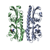
 Movie
Movie Controller


+ Open data

- Basic information
| Entry | Database: PDB / ID: 3irr | ||||||
|---|---|---|---|---|---|---|---|
| Title | Crystal Structure of a Z-Z junction (with HEPES intercalating) | ||||||
 Components Components |
| ||||||
 Keywords Keywords | HYDROLASE/DNA / Z-DNA / ADAR1 / RNA EDITING / INNATE IMMUNITY / DNA JUNCTION / Z DOMAIN / INTERCALATION / Alternative promoter usage / Alternative splicing / Cytoplasm / Disease mutation / DNA-binding / Hydrolase / Isopeptide bond / Metal-binding / mRNA processing / Nucleus / Phosphoprotein / Polymorphism / RNA-binding / RNA-mediated gene silencing / Ubl conjugation / Zinc / HYDROLASE-DNA COMPLEX | ||||||
| Function / homology |  Function and homology information Function and homology informationC6 deamination of adenosine / Formation of editosomes by ADAR proteins / supraspliceosomal complex / double-stranded RNA adenine deaminase / tRNA-specific adenosine deaminase activity / adenosine to inosine editing / negative regulation of protein kinase activity by regulation of protein phosphorylation / double-stranded RNA adenosine deaminase activity / base conversion or substitution editing / response to interferon-alpha ...C6 deamination of adenosine / Formation of editosomes by ADAR proteins / supraspliceosomal complex / double-stranded RNA adenine deaminase / tRNA-specific adenosine deaminase activity / adenosine to inosine editing / negative regulation of protein kinase activity by regulation of protein phosphorylation / double-stranded RNA adenosine deaminase activity / base conversion or substitution editing / response to interferon-alpha / RISC complex assembly / pre-miRNA processing / mRNA modification / RNA processing / positive regulation of viral genome replication / protein export from nucleus / cellular response to virus / protein import into nucleus / PKR-mediated signaling / response to virus / mRNA processing / Interferon alpha/beta signaling / double-stranded RNA binding / defense response to virus / innate immune response / nucleolus / mitochondrion / DNA binding / RNA binding / nucleoplasm / membrane / metal ion binding / nucleus / cytosol / cytoplasm Similarity search - Function | ||||||
| Biological species |  Homo sapiens (human) Homo sapiens (human) | ||||||
| Method |  X-RAY DIFFRACTION / SYNCHROTRON / MOLECULAR REPLACEMENT / Resolution: 2.65 Å X-RAY DIFFRACTION / SYNCHROTRON / MOLECULAR REPLACEMENT / Resolution: 2.65 Å | ||||||
 Authors Authors | Athanasiadis, A. / de Rosa, M. | ||||||
 Citation Citation | Journal: Proc.Natl.Acad.Sci.USA / Year: 2010 Title: Crystal structure of a junction between two Z-DNA helices. Authors: de Rosa, M. / de Sanctis, D. / Rosario, A.L. / Archer, M. / Rich, A. / Athanasiadis, A. / Carrondo, M.A. | ||||||
| History |
|
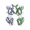
- Structure visualization
| Structure viewer | Molecule: MolmilJmol/JSmol |
|---|
- Downloads & links
-Download
| PDBx/mmCIF format | 3irr.cif.gz | 79.4 KB | Display | PDBx/mmCIF format |
|---|---|---|---|---|
| PDB format | pdb3irr.ent.gz | 56.2 KB | Display | PDB format |
| PDBx/mmJSON format | 3irr.json.gz | Tree view | PDBx/mmJSON format | |
| Others |  Other downloads Other downloads |
-Validation report
| Arichive directory | https://data.pdbj.org/pub/pdb/validation_reports/ir/3irrftp://data.pdbj.org/pub/pdb/validation_reports/ir/3irr | HTTPS FTP |
|---|
-Related structure data
| Related structure data |  3irqC  1qbjS S: Starting model for refinement C: citing same article ( |
|---|---|
| Similar structure data |


-Links
 PDBj
PDBj


- Assembly
| Deposited unit | 
| ||||||||
|---|---|---|---|---|---|---|---|---|---|
| 1 |
| ||||||||
| Unit cell |
| ||||||||
| Details | Biological assembly is a double stranded DNA molecule bound by four protein molecules |
-Components
| #1: Protein | Mass: 7372.584 Da / Num. of mol.: 4 / Fragment: Zalpha domain Source method: isolated from a genetically manipulated source Source: (gene. exp.) Homo sapiens (human) / Gene: ADAR, ADAR1, DSRAD, G1P1, IFI4 / Plasmid: pET28 / Production host:  References: UniProt: P55265, Hydrolases; Acting on carbon-nitrogen bonds, other than peptide bonds; In cyclic amidines #2: DNA chain | | Mass: 4602.959 Da / Num. of mol.: 1 / Source method: obtained synthetically #3: DNA chain | | Mass: 4580.963 Da / Num. of mol.: 1 / Source method: obtained synthetically #4: Chemical | ChemComp-EPE / |   Mass: 238.305 Da / Num. of mol.: 1 / Source method: obtained synthetically / Formula: C8H18N2O4S / Comment: pH buffer*YM Mass: 238.305 Da / Num. of mol.: 1 / Source method: obtained synthetically / Formula: C8H18N2O4S / Comment: pH buffer*YM#5: Water | ChemComp-HOH / |  Mass: 18.015 Da / Num. of mol.: 32 / Source method: isolated from a natural source / Formula: H2O Mass: 18.015 Da / Num. of mol.: 32 / Source method: isolated from a natural source / Formula: H2O |
|---|
-Experimental details
-Experiment
| Experiment | Method: X-RAY DIFFRACTION / Number of used crystals: 1 |
|---|
- Sample preparation
| Crystal | Density Matthews: 2.18 Å3/Da / Density % sol: 43.57 % |
|---|---|
| Crystal grow | Temperature: 293 K / Method: vapor diffusion, hanging drop / pH: 7 Details: 16% PEG 2000 MME, 0.1 M HEPES, 0.2 M ammonium acetate, pH 7.0, VAPOR DIFFUSION, HANGING DROP, temperature 293K |
-Data collection
| Diffraction | Mean temperature: 110 K |
|---|---|
| Diffraction source | Source: SYNCHROTRON / Site: ESRF  / Beamline: ID29 / Wavelength: 0.87 Å / Beamline: ID29 / Wavelength: 0.87 Å |
| Detector | Type: ADSC QUANTUM 315r / Detector: CCD / Date: Jun 27, 2008 |
| Radiation | Monochromator: Si(111) / Protocol: SINGLE WAVELENGTH / Monochromatic (M) / Laue (L): M / Scattering type: x-ray |
| Radiation wavelength | Wavelength: 0.87 Å / Relative weight: 1 |
| Reflection | Resolution: 2.65→47.04 Å / Num. all: 10248 / Num. obs: 10230 / % possible obs: 99.8 % / Observed criterion σ(F): 0 / Observed criterion σ(I): 0 / Redundancy: 4.1 % / Biso Wilson estimate: 64.54 Å2 / Rmerge(I) obs: 0.092 / Rsym value: 0.049 / Net I/σ(I): 12.2 |
| Reflection shell | Resolution: 2.65→2.79 Å / Redundancy: 4.1 % / Rmerge(I) obs: 0.534 / Mean I/σ(I) obs: 2.8 / Num. unique all: 1488 / Rsym value: 0.294 / % possible all: 99.9 |
- Processing
| Software |
| |||||||||||||||||||||||||||||||||||
|---|---|---|---|---|---|---|---|---|---|---|---|---|---|---|---|---|---|---|---|---|---|---|---|---|---|---|---|---|---|---|---|---|---|---|---|---|
| Refinement | Method to determine structure: MOLECULAR REPLACEMENT Starting model: PDB entry 1QBJ Resolution: 2.65→46.023 Å / SU ML: 0.39 / σ(F): 1.36 / σ(I): 0 / Stereochemistry target values: ML
| |||||||||||||||||||||||||||||||||||
| Solvent computation | Shrinkage radii: 0.9 Å / VDW probe radii: 1.11 Å / Solvent model: FLAT BULK SOLVENT MODEL / Bsol: 29.41 Å2 / ksol: 0.324 e/Å3 | |||||||||||||||||||||||||||||||||||
| Refinement step | Cycle: LAST / Resolution: 2.65→46.023 Å
| |||||||||||||||||||||||||||||||||||
| Refine LS restraints |
| |||||||||||||||||||||||||||||||||||
| LS refinement shell |
|
