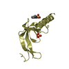
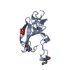
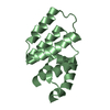
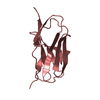
[English] 日本語
Yorodumi
- PDB-3irc: Crystal structure analysis of dengue-1 envelope protein domain III -

+
Open data


ID or keywords:

Loading...

-
Basic information

Entry
Database: PDB / ID: 3irc
TitleCrystal structure analysis of dengue-1 envelope protein domain III
ComponentsENVELOPE PROTEINViral envelope
KeywordsVIRAL PROTEIN / VIRUS / ENVELOPE / STRUCTURAL GENOMICS / CENTER FOR STRUCTURAL GENOMICS OF INFECTIOUS DISEASES / CSGID / ATP-binding / Envelope protein / Helicase / Hydrolase / Membrane / Nucleotide-binding / RNA replication / Transmembrane / Virion
Function / homology
Function and homology information


symbiont-mediated suppression of host JAK-STAT cascade via inhibition of host TYK2 activity / host cell mitochondrion / symbiont-mediated suppression of host JAK-STAT cascade via inhibition of STAT2 activity / symbiont-mediated suppression of host cytoplasmic pattern recognition receptor signaling pathway via inhibition of MAVS activity / ribonucleoside triphosphate phosphatase activity / viral capsid / : / double-stranded RNA binding / protein complex oligomerization / monoatomic ion channel activity ...symbiont-mediated suppression of host JAK-STAT cascade via inhibition of host TYK2 activity / host cell mitochondrion / symbiont-mediated suppression of host JAK-STAT cascade via inhibition of STAT2 activity / symbiont-mediated suppression of host cytoplasmic pattern recognition receptor signaling pathway via inhibition of MAVS activity / ribonucleoside triphosphate phosphatase activity / viral capsid / : / double-stranded RNA binding / protein complex oligomerization / monoatomic ion channel activity / clathrin-dependent endocytosis of virus by host cell / mRNA (nucleoside-2'-O-)-methyltransferase activity / mRNA 5'-cap (guanine-N7-)-methyltransferase activity / RNA helicase activity / host cell endoplasmic reticulum membrane / protein dimerization activity / induction by virus of host autophagy / viral RNA genome replication / RNA-dependent RNA polymerase activity / serine-type endopeptidase activity / fusion of virus membrane with host endosome membrane / viral envelope / symbiont-mediated suppression of host type I interferon-mediated signaling pathway / host cell nucleus / virion attachment to host cell / virion membrane / structural molecule activity / proteolysis / extracellular region / ATP binding / membrane / metal ion binding
Similarity search - Function
Immunoglobulin-like - #350 / : / Flavivirus envelope glycoprotein E, stem/anchor domain / RNA-directed RNA polymerase, thumb domain, Flavivirus / Flavivirus RNA-directed RNA polymerase, thumb domain / Flavivirus capsid protein C superfamily / : / Flavivirus non-structural protein NS2B / Flavivirus NS3 helicase, C-terminal helical domain / Genome polyprotein, Flavivirus ...Immunoglobulin-like - #350 / : / Flavivirus envelope glycoprotein E, stem/anchor domain / RNA-directed RNA polymerase, thumb domain, Flavivirus / Flavivirus RNA-directed RNA polymerase, thumb domain / Flavivirus capsid protein C superfamily / : / Flavivirus non-structural protein NS2B / Flavivirus NS3 helicase, C-terminal helical domain / Genome polyprotein, Flavivirus / Flavivirus non-structural protein NS4A / Flavivirus non-structural protein NS2B / Flavivirus capsid protein C / Flavivirus non-structural protein NS4B / mRNA cap 0/1 methyltransferase / Flavivirus capsid protein C / Flavivirus non-structural protein NS4B / Flavivirus non-structural protein NS4A / Flavivirus NS2B domain profile. / mRNA cap 0 and cap 1 methyltransferase (EC 2.1.1.56 and EC 2.1.1.57) domain profile. / Flavivirus non-structural protein NS2A / Flavivirus non-structural protein NS2A / Flavivirus NS3, petidase S7 / Peptidase S7, Flavivirus NS3 serine protease / Flavivirus NS3 protease (NS3pro) domain profile. / Envelope glycoprotein M, flavivirus / Flavivirus envelope glycoprotein M / RNA-directed RNA polymerase, flavivirus / Flavivirus RNA-directed RNA polymerase, fingers and palm domains / Flavivirus non-structural Protein NS1 / Flavivirus non-structural protein NS1 / Envelope glycoprotein M superfamily, flavivirus / Flavivirus polyprotein propeptide / Flavivirus polyprotein propeptide superfamily / Flavivirus polyprotein propeptide / Flaviviral glycoprotein E, central domain, subdomain 1 / Flaviviral glycoprotein E, central domain, subdomain 2 / Flavivirus envelope glycoprotein E, Stem/Anchor domain / Flavivirus glycoprotein E, immunoglobulin-like domain / Flavivirus envelope glycoprotein E, Stem/Anchor domain superfamily / Flavivirus glycoprotein, immunoglobulin-like domain / Flavivirus glycoprotein central and dimerisation domain / Flavivirus glycoprotein, central and dimerisation domains / Ribosomal RNA methyltransferase, FtsJ domain / FtsJ-like methyltransferase / Flavivirus/Alphavirus glycoprotein, immunoglobulin-like domain superfamily / Flavivirus glycoprotein, central and dimerisation domain superfamily / Flaviviral glycoprotein E, dimerisation domain / DEAD box, Flavivirus / Flavivirus DEAD domain / helicase superfamily c-terminal domain / Immunoglobulin E-set / Superfamilies 1 and 2 helicase C-terminal domain profile. / Superfamilies 1 and 2 helicase ATP-binding type-1 domain profile. / DEAD-like helicases superfamily / Helicase, C-terminal / Helicase superfamily 1/2, ATP-binding domain / RNA-directed RNA polymerase, catalytic domain / RdRp of positive ssRNA viruses catalytic domain profile. / S-adenosyl-L-methionine-dependent methyltransferase superfamily / Peptidase S1, PA clan, chymotrypsin-like fold / Peptidase S1, PA clan / DNA/RNA polymerase superfamily / Immunoglobulin-like / Sandwich / P-loop containing nucleoside triphosphate hydrolase / Mainly Beta
Similarity search - Domain/homology
Biological speciesDengue virus 1
MethodX-RAY DIFFRACTION / SYNCHROTRON / MOLECULAR REPLACEMENT / molecular replacement / Resolution: 2.25 Å
AuthorsNelson, C.A. / Kim, T. / Warren, J.T. / Chruszcz, M. / Minor, W. / Fremont, D.H. / Center for Structural Genomics of Infectious Diseases (CSGID)
CitationJournal: To be Published
Title: Crystal Structure Analysis of the Dengue-1 Envelope Protein Domain III
Authors: Nelson, C.A. / Kim, T. / Warren, J.T. / Fremont, D.H.
History
DepositionAug 21, 2009Deposition site: RCSB / Processing site: RCSB
SupersessionSep 29, 2009ID: 3EGP
Revision 1.0Sep 29, 2009Provider: repository / Type: Initial release
Revision 1.1Jul 13, 2011Group: Advisory / Version format compliance
Revision 1.2Nov 1, 2017Group: Advisory / Refinement description / Category: pdbx_unobs_or_zero_occ_residues / software
Item: _software.classification / _software.contact_author ..._software.classification / _software.contact_author / _software.contact_author_email / _software.date / _software.language / _software.location / _software.name / _software.type / _software.version
Revision 1.3Apr 13, 2022Group: Advisory / Database references ...Advisory / Database references / Derived calculations / Structure summary
Category: audit_author / database_2 ...audit_author / database_2 / pdbx_unobs_or_zero_occ_residues / struct_ref_seq_dif / struct_site
Item: _audit_author.identifier_ORCID / _database_2.pdbx_DOI ..._audit_author.identifier_ORCID / _database_2.pdbx_DOI / _database_2.pdbx_database_accession / _struct_ref_seq_dif.details / _struct_site.pdbx_auth_asym_id / _struct_site.pdbx_auth_comp_id / _struct_site.pdbx_auth_seq_id
Revision 1.4Sep 6, 2023Group: Data collection / Refinement description
Category: chem_comp_atom / chem_comp_bond / pdbx_initial_refinement_model

-
Structure visualization

Structure viewerMolecule:
MolmilJmol/JSmol

Downloads & links

-
Assembly

Deposited unit
A: ENVELOPE PROTEIN
hetero molecules


Theoretical massNumber of molelcules
Total (without water)12,0815
Polymers11,6961
Non-polymers3844
Water95553
1


  • Idetical with deposited unit
  • defined by author
TypeNameSymmetry operationNumber
identity operation1_555x,y,z1
2
A: ENVELOPE PROTEIN
hetero molecules

A: ENVELOPE PROTEIN
hetero molecules

A: ENVELOPE PROTEIN
hetero molecules


Theoretical massNumber of molelcules
Total (without water)36,24215
Polymers35,0893
Non-polymers1,15312
Water543
TypeNameSymmetry operationNumber
identity operation1_555x,y,z1
crystal symmetry operation5_555z,x,y1
crystal symmetry operation9_555y,z,x1
Buried area3010 Å2
ΔGint-25 kcal/mol
Surface area16560 Å2
MethodPISA
Unit cell
Length a, b, c (Å)88.087, 88.087, 88.087
Angle α, β, γ (deg.)90.00, 90.00, 90.00
Int Tables number198
Space group name H-MP213
Components on special symmetry positions
IDModelComponents
11A-1-

SO4

-
Components

#1: Protein ENVELOPE PROTEIN / Viral envelope


Mass: 11696.393 Da / Num. of mol.: 1 / Fragment: DOMAIN III, residues 576-679
Source method: isolated from a genetically manipulated source
Source: (gene. exp.) Dengue virus 1 / Strain: 16007 / Gene: ENVELOPE / Plasmid: pET21 / Production host: Escherichia coli (E. coli) / Strain (production host): BL21-CodonPlus(DE3)-RIL / References: UniProt: Q9J7C6
#2: Chemical
ChemComp-SO4 / SULFATE ION / Sulfate


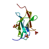
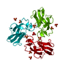
Mass: 96.063 Da / Num. of mol.: 4 / Source method: obtained synthetically / Formula: SO4
#3: Water ChemComp-HOH / water / Water


Mass: 18.015 Da / Num. of mol.: 53 / Source method: isolated from a natural source / Formula: H2O

-
Experimental details

-
Experiment

ExperimentMethod: X-RAY DIFFRACTION / Number of used crystals: 1

-
Sample preparation

CrystalDensity Matthews: 5.076 Å3/Da / Density % sol: 75.77 %
Crystal growTemperature: 293 K / Method: vapor diffusion, hanging drop / pH: 7.5
Details: 0.1M IMIDAZOLE, 0.2M LITHIUM SULFATE, 30% PEG 3000, PH 7.5, VAPOR DIFFUSION, HANGING DROP, TEMPERATURE 293K

-
Data collection

DiffractionMean temperature: 100 K
Diffraction sourceSource: SYNCHROTRON / Site: ALS / Beamline: 4.2.2 / Wavelength: 1.239 Å
DetectorType: NOIR-1 / Detector: CCD
RadiationProtocol: SINGLE WAVELENGTH / Monochromatic (M) / Laue (L): M / Scattering type: x-ray
Radiation wavelengthWavelength: 1.239 Å / Relative weight: 1
ReflectionResolution: 2.2→35.97 Å / Num. obs: 10458 / % possible obs: 99.3 % / Redundancy: 3.8 % / Rmerge(I) obs: 0.039 / Rsym value: 0.038 / Net I/σ(I): 17.7
Reflection shellResolution: 2.25→2.29 Å / Redundancy: 3.3 % / Rmerge(I) obs: 0.694 / Mean I/σ(I) obs: 1.92 / Rsym value: 0.627 / % possible all: 98.5

-
Phasing

PhasingMethod: molecular replacement

-
Processing

Software
NameVersionClassificationNB
DENZOdata reduction
SCALEPACKdata scaling
PHASERphasing
REFMAC5.5.0091refinement
PDB_EXTRACT3.005data extraction
HKL-2000data reduction
RefinementMethod to determine structure: MOLECULAR REPLACEMENT
Starting model: 1OAN
Resolution: 2.25→35.97 Å / Cor.coef. Fo:Fc: 0.956 / Cor.coef. Fo:Fc free: 0.946 / Occupancy max: 1 / Occupancy min: 0 / SU B: 8.794 / SU ML: 0.098 / TLS residual ADP flag: LIKELY RESIDUAL / Cross valid method: THROUGHOUT / ESU R: 0.165 / ESU R Free: 0.143 / Stereochemistry target values: MAXIMUM LIKELIHOOD / Details: HYDROGENS HAVE BEEN ADDED IN THE RIDING POSITIONS
RfactorNum. reflection% reflectionSelection details
Rfree0.22144 520 4.7 %RANDOM
Rwork0.21145 ---
obs0.21193 10458 99.09 %-
Solvent computationIon probe radii: 0.8 Å / Shrinkage radii: 0.8 Å / VDW probe radii: 1.4 Å / Solvent model: MASK
Displacement parametersBiso mean: 32.821 Å2
Refinement stepCycle: LAST / Resolution: 2.25→35.97 Å
ProteinNucleic acidLigandSolventTotal
Num. atoms787 0 20 53 860
Refine LS restraints
Refine-IDTypeDev idealDev ideal targetNumber
X-RAY DIFFRACTIONr_bond_refined_d0.0170.022783
X-RAY DIFFRACTIONr_bond_other_d
X-RAY DIFFRACTIONr_angle_refined_deg1.812.0011061
X-RAY DIFFRACTIONr_angle_other_deg
X-RAY DIFFRACTIONr_dihedral_angle_1_deg7.326598
X-RAY DIFFRACTIONr_dihedral_angle_2_deg34.60226.07128
X-RAY DIFFRACTIONr_dihedral_angle_3_deg14.44515137
X-RAY DIFFRACTIONr_dihedral_angle_4_deg37.329151
X-RAY DIFFRACTIONr_chiral_restr0.0940.2116
X-RAY DIFFRACTIONr_gen_planes_refined0.010.021565
X-RAY DIFFRACTIONr_gen_planes_other
X-RAY DIFFRACTIONr_nbd_refined
X-RAY DIFFRACTIONr_nbd_other
X-RAY DIFFRACTIONr_nbtor_refined
X-RAY DIFFRACTIONr_nbtor_other
X-RAY DIFFRACTIONr_xyhbond_nbd_refined
X-RAY DIFFRACTIONr_xyhbond_nbd_other
X-RAY DIFFRACTIONr_metal_ion_refined
X-RAY DIFFRACTIONr_metal_ion_other
X-RAY DIFFRACTIONr_symmetry_vdw_refined
X-RAY DIFFRACTIONr_symmetry_vdw_other
X-RAY DIFFRACTIONr_symmetry_hbond_refined
X-RAY DIFFRACTIONr_symmetry_hbond_other
X-RAY DIFFRACTIONr_symmetry_metal_ion_refined
X-RAY DIFFRACTIONr_symmetry_metal_ion_other
X-RAY DIFFRACTIONr_mcbond_it1.7791.5491
X-RAY DIFFRACTIONr_mcbond_other
X-RAY DIFFRACTIONr_mcangle_it2.9112795
X-RAY DIFFRACTIONr_scbond_it4.6773292
X-RAY DIFFRACTIONr_scangle_it7.1164.5266
X-RAY DIFFRACTIONr_rigid_bond_restr
X-RAY DIFFRACTIONr_sphericity_free
X-RAY DIFFRACTIONr_sphericity_bonded
LS refinement shellResolution: 2.25→2.309 Å / Total num. of bins used: 20
RfactorNum. reflection% reflection
Rfree0.388 35 -
Rwork0.332 766 -
obs--98.52 %
Refinement TLS params.

Method: refined / Refine-ID: X-RAY DIFFRACTION

IDL112)L122)L132)L222)L232)L332)S11 (Å °)S12 (Å °)S13 (Å °)S21 (Å °)S22 (Å °)S23 (Å °)S31 (Å °)S32 (Å °)S33 (Å °)T112)T122)T132)T222)T232)T332)Origin x (Å)Origin y (Å)Origin z (Å)
18.8128-1.0833-5.73163.87860.13846.09620.18980.2450.5461-0.13940.03330.12-0.069-0.2516-0.22310.21380.07990.02550.2680.10340.2607-24.726-3.207-12.797
213.78020.6821-7.16154.54-1.871811.7138-0.2950.7328-0.1587-0.63440.0053-0.38960.5057-0.65760.28970.18190.06940.00250.25990.03140.1411-26.011-5.539-15.736
310.87932.3759-4.63132.6263-1.38584.82630.1130.10160.19360.1522-0.0119-0.08870.3216-0.1396-0.10120.23730.03810.03470.0840.02340.1789-33.75-7.18-4.447
Refinement TLS group
IDRefine-IDRefine TLS-IDAuth asym-IDAuth seq-ID
1X-RAY DIFFRACTION1A296 - 343
2X-RAY DIFFRACTION2A344 - 365
3X-RAY DIFFRACTION3A366 - 399

+
About Yorodumi

-
News

-
Feb 9, 2022. New format data for meta-information of EMDB entries

New format data for meta-information of EMDB entries

  • Version 3 of the EMDB header file is now the official format.
  • The previous official version 1.9 will be removed from the archive.

Related info.:EMDB header

External links:wwPDB to switch to version 3 of the EMDB data model

-
Aug 12, 2020. Covid-19 info

Covid-19 info

URL: https://pdbj.org/emnavi/covid19.php

New page: Covid-19 featured information page in EM Navigator.

Related info.:Covid-19 info / Mar 5, 2020. Novel coronavirus structure data

+
Mar 5, 2020. Novel coronavirus structure data

Novel coronavirus structure data

Related info.:Yorodumi Speices / Aug 12, 2020. Covid-19 info

External links:COVID-19 featured content - PDBj / Molecule of the Month (242):Coronavirus Proteases

+
Jan 31, 2019. EMDB accession codes are about to change! (news from PDBe EMDB page)

EMDB accession codes are about to change! (news from PDBe EMDB page)

  • The allocation of 4 digits for EMDB accession codes will soon come to an end. Whilst these codes will remain in use, new EMDB accession codes will include an additional digit and will expand incrementally as the available range of codes is exhausted. The current 4-digit format prefixed with “EMD-” (i.e. EMD-XXXX) will advance to a 5-digit format (i.e. EMD-XXXXX), and so on. It is currently estimated that the 4-digit codes will be depleted around Spring 2019, at which point the 5-digit format will come into force.
  • The EM Navigator/Yorodumi systems omit the EMD- prefix.

Related info.:Q: What is EMD? / ID/Accession-code notation in Yorodumi/EM Navigator

External links:EMDB Accession Codes are Changing Soon! / Contact to PDBj

+
Jul 12, 2017. Major update of PDB

Major update of PDB

  • wwPDB released updated PDB data conforming to the new PDBx/mmCIF dictionary.
  • This is a major update changing the version number from 4 to 5, and with Remediation, in which all the entries are updated.
  • In this update, many items about electron microscopy experimental information are reorganized (e.g. em_software).
  • Now, EM Navigator and Yorodumi are based on the updated data.

External links:wwPDB Remediation / Enriched Model Files Conforming to OneDep Data Standards Now Available in the PDB FTP Archive

-
Yorodumi

Thousand views of thousand structures

  • Yorodumi is a browser for structure data from EMDB, PDB, SASBDB, etc.
  • This page is also the successor to EM Navigator detail page, and also detail information page/front-end page for Omokage search.
  • The word "yorodu" (or yorozu) is an old Japanese word meaning "ten thousand". "mi" (miru) is to see.

Related info.:EMDB / PDB / SASBDB / Comparison of 3 databanks / Yorodumi Search / Aug 31, 2016. New EM Navigator & Yorodumi / Yorodumi Papers / Jmol/JSmol / Function and homology information / Changes in new EM Navigator and Yorodumi

Read more