- PDB-3ipv: Crystal structure of Spatholobus parviflorus seed lectin -
+
Open data
ID or keywords:
Loading...
-
Basic information
Entry
Database: PDB / ID: 3ipv
Title
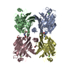
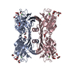
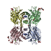
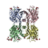
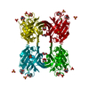
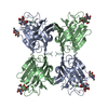
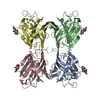
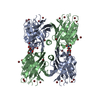
Crystal structure of Spatholobus parviflorus seed lectin
Components
Lectin alpha chain
Lectin beta chain
Keywords
SUGAR BINDING PROTEIN / Galactose binding / Seed lectin / Hemagglutinin / Legume lectin / Anti fungal
Function / homology
Function and homology information
defense response to fungus / manganese ion binding / carbohydrate binding / killing of cells of another organism / calcium ion binding / protein-containing complex Similarity search - Function
Mass: 18.015 Da / Num. of mol.: 693 / Source method: isolated from a natural source / Formula: H2O
Sequence details
THE DEPOSITOR GUIDED BY THE SEQUENCE OF THE MODEL (OF MOLECULAR REPLACEMENT)POLYPEPTIDE. THE ...THE DEPOSITOR GUIDED BY THE SEQUENCE OF THE MODEL (OF MOLECULAR REPLACEMENT)POLYPEPTIDE. THE AMBIGUITY AT CERTAIN PLACES IN INTERPRETING ELECTRON DENSITY (E.G. ASP/ASN, ETC.) FOR SEQUENCE CAN NOT BE RULED OUT. A BREAK IS FOUND IN THE ELECTRON DENSITY MAP CALCULATED USING THE X-RAY DIFFRACTION DATA. IN THE CASE OF SPATHOLOBUS PARVIFLORUS ALPHA CHAIN, THE BREAK FOUND MAY ACCOMMODATE A MINIMUM OF TWO RESIDUES. HENCE, THE ABOVE SEQUENCE IS PROVIDED AS UNK (240, 241).
-
Experimental details
-
Experiment
Experiment
Method: X-RAY DIFFRACTION / Number of used crystals: 1
-
Sample preparation
Crystal
Density Matthews: 2.66 Å3/Da / Density % sol: 53.78 %
Crystal grow
Temperature: 298 K / Method: vapor diffusion, hanging drop / pH: 7.4 Details: 25% PEG 8000, 0.2M Phosphate Buffer Saline, 5% MPD, 5% Iso propanol, pH 7.4, VAPOR DIFFUSION, HANGING DROP, temperature 298K
-
Data collection
Diffraction
Mean temperature: 100 K
Diffraction source
Source: ROTATING ANODE / Type: BRUKER AXS MICROSTAR / Wavelength: 1.5418 Å
Detector
Type: MAR scanner 345 mm plate / Detector: IMAGE PLATE / Date: Mar 12, 2009 / Details: MIRROR
Radiation
Monochromator: OSMIC MIRROR / Protocol: SINGLE WAVELENGTH / Monochromatic (M) / Laue (L): M / Scattering type: x-ray
In the structure databanks used in Yorodumi, some data are registered as the other names, "COVID-19 virus" and "2019-nCoV". Here are the details of the virus and the list of structure data.
Jan 31, 2019. EMDB accession codes are about to change! (news from PDBe EMDB page)
EMDB accession codes are about to change! (news from PDBe EMDB page)
The allocation of 4 digits for EMDB accession codes will soon come to an end. Whilst these codes will remain in use, new EMDB accession codes will include an additional digit and will expand incrementally as the available range of codes is exhausted. The current 4-digit format prefixed with “EMD-” (i.e. EMD-XXXX) will advance to a 5-digit format (i.e. EMD-XXXXX), and so on. It is currently estimated that the 4-digit codes will be depleted around Spring 2019, at which point the 5-digit format will come into force.
The EM Navigator/Yorodumi systems omit the EMD- prefix.
Related info.:Q: What is EMD? / ID/Accession-code notation in Yorodumi/EM Navigator
Yorodumi is a browser for structure data from EMDB, PDB, SASBDB, etc.
This page is also the successor to EM Navigator detail page, and also detail information page/front-end page for Omokage search.
The word "yorodu" (or yorozu) is an old Japanese word meaning "ten thousand". "mi" (miru) is to see.
Related info.:EMDB / PDB / SASBDB / Comparison of 3 databanks / Yorodumi Search / Aug 31, 2016. New EM Navigator & Yorodumi / Yorodumi Papers / Jmol/JSmol / Function and homology information / Changes in new EM Navigator and Yorodumi
 Movie
Movie Controller


 Open data
 Basic information
 Components
 Keywords
 Function and homology information
 Spatholobus parviflorus (plant)
 X-RAY DIFFRACTION /
  Authors
 Citation
 Structure visualization
 Downloads & links
 Other downloads
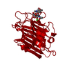


 PDBj
PDBj


 Assembly
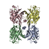

 Mass: 40.078 Da / Num. of mol.: 4 / Source method: obtained synthetically / Formula: Ca
 Mass: 54.938 Da / Num. of mol.: 4 / Source method: obtained synthetically / Formula: Mn
 Mass: 18.015 Da / Num. of mol.: 693 / Source method: isolated from a natural source / Formula: H2O
 Sample preparation
 Processing