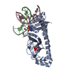
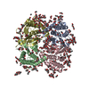
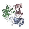
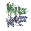
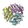
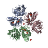
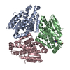
- PDB-3io5: Crystal Structure of a dimeric form of the uvsX Recombinase core ... -
+
Open data
ID or keywords:
Loading...
-
Basic information
Entry
Database: PDB / ID: 3io5
Title
Crystal Structure of a dimeric form of the uvsX Recombinase core domain from Enterobacteria Phage T4
Components
Recombination and repair protein
Keywords
DNA BINDING PROTEIN / storage dimer / inactive conformation / RecA like core domain / ATP-binding / DNA damage / DNA recombination / DNA repair / DNA replication / Nucleotide-binding
Function / homology
Function and homology information
ATP-dependent DNA damage sensor activity / single-stranded DNA binding / DNA recombination / DNA replication / DNA repair / ATP binding Similarity search - Function
: / Bacteriophage T4, Recombination and repair protein, C-terminal domain / DNA recombination and repair protein RecA / : / RecA N-terminal domain / DNA recombination and repair protein RecA, monomer-monomer interface / RecA family profile 2. / DNA recombination and repair protein RecA-like, ATP-binding domain / RecA family profile 1. / P-loop containing nucleotide triphosphate hydrolases ...: / Bacteriophage T4, Recombination and repair protein, C-terminal domain / DNA recombination and repair protein RecA / : / RecA N-terminal domain / DNA recombination and repair protein RecA, monomer-monomer interface / RecA family profile 2. / DNA recombination and repair protein RecA-like, ATP-binding domain / RecA family profile 1. / P-loop containing nucleotide triphosphate hydrolases / Rossmann fold / P-loop containing nucleoside triphosphate hydrolase / 3-Layer(aba) Sandwich / Alpha Beta Similarity search - Domain/homology
Resolution: 2.35→2.462 Å / Redundancy: 5.4 % / Mean I/σ(I) obs: 3.7 / Num. unique all: 2394 / Rsym value: 0.345 / % possible all: 84.7
-
Processing
Software
Name
Version
Classification
NB
DENZO
datareduction
SCALEPACK
datascaling
REFMAC
5.5.0035
refinement
PDB_EXTRACT
3.005
dataextraction
MAR345dtb
datacollection
HKL-2000
datareduction
HKL-2000
datascaling
PHENIX
phasing
Refinement
Method to determine structure: SAD / Resolution: 2.4→41.56 Å / Cor.coef. Fo:Fc: 0.945 / Cor.coef. Fo:Fc free: 0.921 / Occupancy max: 1 / Occupancy min: 1 / SU B: 18.557 / SU ML: 0.191 / Cross valid method: THROUGHOUT / σ(F): 0 / ESU R: 0.374 / ESU R Free: 0.251 / Stereochemistry target values: MAXIMUM LIKELIHOOD Details: HYDROGENS HAVE BEEN ADDED IN THE RIDING POSITIONS U VALUES: RESIDUAL ONLY
Rfactor
Num. reflection
% reflection
Selection details
Rfree
0.248
1330
5 %
RANDOM
Rwork
0.21
-
-
-
obs
0.212
26592
99.25 %
-
Solvent computation
Ion probe radii: 0.8 Å / Shrinkage radii: 0.8 Å / VDW probe radii: 1.2 Å / Solvent model: MASK
In the structure databanks used in Yorodumi, some data are registered as the other names, "COVID-19 virus" and "2019-nCoV". Here are the details of the virus and the list of structure data.
Jan 31, 2019. EMDB accession codes are about to change! (news from PDBe EMDB page)
EMDB accession codes are about to change! (news from PDBe EMDB page)
The allocation of 4 digits for EMDB accession codes will soon come to an end. Whilst these codes will remain in use, new EMDB accession codes will include an additional digit and will expand incrementally as the available range of codes is exhausted. The current 4-digit format prefixed with “EMD-” (i.e. EMD-XXXX) will advance to a 5-digit format (i.e. EMD-XXXXX), and so on. It is currently estimated that the 4-digit codes will be depleted around Spring 2019, at which point the 5-digit format will come into force.
The EM Navigator/Yorodumi systems omit the EMD- prefix.
Related info.:Q: What is EMD? / ID/Accession-code notation in Yorodumi/EM Navigator
Yorodumi is a browser for structure data from EMDB, PDB, SASBDB, etc.
This page is also the successor to EM Navigator detail page, and also detail information page/front-end page for Omokage search.
The word "yorodu" (or yorozu) is an old Japanese word meaning "ten thousand". "mi" (miru) is to see.
Related info.:EMDB / PDB / SASBDB / Comparison of 3 databanks / Yorodumi Search / Aug 31, 2016. New EM Navigator & Yorodumi / Yorodumi Papers / Jmol/JSmol / Function and homology information / Changes in new EM Navigator and Yorodumi
 Movie
Movie Controller

 Yorodumi
 Open data
 Basic information
 Components
 Keywords
 Function and homology information
 Enterobacteria phage T4 (virus)
 X-RAY DIFFRACTION /
  Authors
 Citation
 Structure visualization
 Downloads & links
 Other downloads
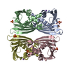


 PDBj
PDBj
 Assembly


 Mass: 94.971 Da / Num. of mol.: 2 / Source method: obtained synthetically / Formula: PO4
 Mass: 18.015 Da / Num. of mol.: 55 / Source method: isolated from a natural source / Formula: H2O
 Sample preparation
 / Beamline: 22-ID / Wavelength: 1 Å
 Processing