 Movie
Movie Controller

[English] 日本語
 Yorodumi
- PDB-1sg4: Crystal structure of human mitochondrial delta3-delta2-enoyl-CoA ... -
+ Open data

- Basic information
| Entry | Database: PDB / ID: 1sg4 | ||||||
|---|---|---|---|---|---|---|---|
| Title | Crystal structure of human mitochondrial delta3-delta2-enoyl-CoA isomerase | ||||||
 Components Components | 3,2-trans-enoyl-CoA isomerase, mitochondrial | ||||||
 Keywords Keywords | ISOMERASE / crotonase fold | ||||||
| Function / homology |  Function and homology information Function and homology informationmitochondrial fatty acid beta-oxidation of unsaturated fatty acids / intramolecular oxidoreductase activity, transposing C=C bonds / Delta3-Delta2-enoyl-CoA isomerase / delta(3)-delta(2)-enoyl-CoA isomerase activity / fatty acid beta-oxidation / Mitochondrial protein degradation / mitochondrial matrix / mitochondrion Similarity search - Function | ||||||
| Biological species |  Homo sapiens (human) Homo sapiens (human) | ||||||
| Method |  X-RAY DIFFRACTION / SYNCHROTRON / MAD / Resolution: 1.3 Å X-RAY DIFFRACTION / SYNCHROTRON / MAD / Resolution: 1.3 Å | ||||||
 Authors Authors | Partanen, S.T. / Novikov, D.K. / Popov, A.N. / Mursula, A.M. / Hiltunen, J.K. / Wierenga, R.K. | ||||||
 Citation Citation | Journal: J.Mol.Biol. / Year: 2004 Title: The 1.3 A crystal structure of human mitochondrial Delta3-Delta2-enoyl-CoA isomerase shows a novel mode of binding for the fatty acyl group. Authors: Partanen, S.T. / Novikov, D.K. / Popov, A.N. / Mursula, A.M. / Hiltunen, J.K. / Wierenga, R.K. | ||||||
| History |
|
- Structure visualization
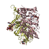
| Structure viewer | Molecule: MolmilJmol/JSmol |
|---|
- Downloads & links
-Download
| PDBx/mmCIF format | 1sg4.cif.gz | 308.9 KB | Display | PDBx/mmCIF format |
|---|---|---|---|---|
| PDB format | pdb1sg4.ent.gz | 254.2 KB | Display | PDB format |
| PDBx/mmJSON format | 1sg4.json.gz | Tree view | PDBx/mmJSON format | |
| Others |  Other downloads Other downloads |
-Validation report
| Arichive directory | https://data.pdbj.org/pub/pdb/validation_reports/sg/1sg4ftp://data.pdbj.org/pub/pdb/validation_reports/sg/1sg4 | HTTPS FTP |
|---|
-Related structure data
| Similar structure data |
|---|


-Links
 PDBj
PDBj- Assembly
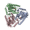
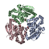
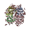
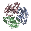
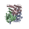
| Deposited unit | 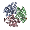
| ||||||||
|---|---|---|---|---|---|---|---|---|---|
| 1 |
| ||||||||
| 2 | 
| ||||||||
| Unit cell |
|
-Components
| #1: Protein | Mass: 28617.965 Da / Num. of mol.: 3 Source method: isolated from a genetically manipulated source Source: (gene. exp.) Homo sapiens (human) / Gene: DCI / Plasmid: pET3a / Production host:  References: UniProt: P42126, Delta3-Delta2-enoyl-CoA isomerase #2: Chemical | ChemComp-CO8 / | 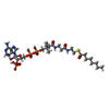  Mass: 893.730 Da / Num. of mol.: 1 / Source method: obtained synthetically / Formula: C29H50N7O17P3S Mass: 893.730 Da / Num. of mol.: 1 / Source method: obtained synthetically / Formula: C29H50N7O17P3S#3: Water | ChemComp-HOH / |  Mass: 18.015 Da / Num. of mol.: 666 / Source method: isolated from a natural source / Formula: H2O Mass: 18.015 Da / Num. of mol.: 666 / Source method: isolated from a natural source / Formula: H2O |
|---|
-Experimental details
-Experiment
| Experiment | Method: X-RAY DIFFRACTION / Number of used crystals: 2 |
|---|
- Sample preparation
| Crystal | Density Matthews: 2.33 Å3/Da / Density % sol: 46.9 % |
|---|---|
| Crystal grow | Temperature: 294.15 K / Method: vapor diffusion, hanging drop / pH: 4.1 Details: PEG 6000, sodium acetate, sodium cloride, pH 4.1, VAPOR DIFFUSION, HANGING DROP, temperature 294.15K |
-Data collection
| Diffraction |
| ||||||||||||||||||
|---|---|---|---|---|---|---|---|---|---|---|---|---|---|---|---|---|---|---|---|
| Diffraction source |
| ||||||||||||||||||
| Detector |
| ||||||||||||||||||
| Radiation |
| ||||||||||||||||||
| Radiation wavelength |
| ||||||||||||||||||
| Reflection | Resolution: 1.3→20 Å / Num. obs: 182696 / % possible obs: 94.5 % / Observed criterion σ(F): 0 / Observed criterion σ(I): 0 / Redundancy: 3.1 % / Biso Wilson estimate: 16.6 Å2 / Rmerge(I) obs: 0.057 / Net I/σ(I): 17.3 | ||||||||||||||||||
| Reflection shell | Resolution: 1.3→1.31 Å / Redundancy: 3.1 % / Rmerge(I) obs: 0.201 / Mean I/σ(I) obs: 2.7 / Num. unique all: 5263 / % possible all: 82.1 |

- Processing
| Software |
| ||||||||||||||||||||
|---|---|---|---|---|---|---|---|---|---|---|---|---|---|---|---|---|---|---|---|---|---|
| Refinement | Method to determine structure: MAD / Resolution: 1.3→20 Å / σ(F): 0 / σ(I): 0 / Stereochemistry target values: Engh & Huber
| ||||||||||||||||||||
| Displacement parameters | Biso mean: 23.8 Å2 | ||||||||||||||||||||
| Refinement step | Cycle: LAST / Resolution: 1.3→20 Å
| ||||||||||||||||||||
| Refine LS restraints |
| ||||||||||||||||||||
| LS refinement shell |
|
