 Movie
Movie Controller


+ Open data

- Basic information
| Entry | Database: PDB / ID: 6cj6 | ||||||
|---|---|---|---|---|---|---|---|
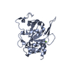
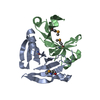
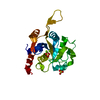
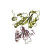
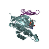
| Title | Structure of the poxvirus protein F9 | ||||||
 Components Components | Protein F9 | ||||||
 Keywords Keywords | VIRAL PROTEIN / entry fusion complex associated protein | ||||||
| Function / homology |  Function and homology information Function and homology information | ||||||
| Biological species |  Vaccinia virus Vaccinia virus | ||||||
| Method |  X-RAY DIFFRACTION / SYNCHROTRON / SAD / Resolution: 2.1 Å X-RAY DIFFRACTION / SYNCHROTRON / SAD / Resolution: 2.1 Å | ||||||
 Authors Authors | Diesterbeck, U.S. / Gittis, A.G. / Garboczi, D.N. / Moss, B. | ||||||
 Citation Citation | Journal: Sci Rep / Year: 2018 Title: The 2.1 angstrom structure of protein F9 and its comparison to L1, two components of the conserved poxvirus entry-fusion complex. Authors: Diesterbeck, U.S. / Gittis, A.G. / Garboczi, D.N. / Moss, B. | ||||||
| History |
|
- Structure visualization
| Structure viewer | Molecule: MolmilJmol/JSmol |
|---|
- Downloads & links
-Download
| PDBx/mmCIF format | 6cj6.cif.gz | 174 KB | Display | PDBx/mmCIF format |
|---|---|---|---|---|
| PDB format | pdb6cj6.ent.gz | 137 KB | Display | PDB format |
| PDBx/mmJSON format | 6cj6.json.gz | Tree view | PDBx/mmJSON format | |
| Others |  Other downloads Other downloads |
-Validation report
| Arichive directory | https://data.pdbj.org/pub/pdb/validation_reports/cj/6cj6ftp://data.pdbj.org/pub/pdb/validation_reports/cj/6cj6 | HTTPS FTP |
|---|
-Related structure data
| Similar structure data |
|---|


-Links
 PDBj
PDBj


- Assembly
| Deposited unit | 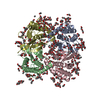
| ||||||||
|---|---|---|---|---|---|---|---|---|---|
| 1 | 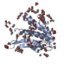
| ||||||||
| 2 | 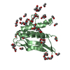
| ||||||||
| 3 | 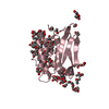
| ||||||||
| 4 | 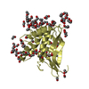
| ||||||||
| Unit cell |
|
-Components
-Protein , 1 types, 4 molecules ABCD
| #1: Protein | Mass: 19635.631 Da / Num. of mol.: 4 / Fragment: residues 1-176 Source method: isolated from a genetically manipulated source Source: (gene. exp.) Vaccinia virus (strain Western Reserve)Strain: Western Reserve / Gene: VACWR048, F9L / Production host:  |
|---|
-Non-polymers , 15 types, 395 molecules 


| #2: Chemical | ChemComp-PG4 /  Mass: 194.226 Da / Num. of mol.: 5 / Source method: obtained synthetically / Formula: C8H18O5 / Comment: precipitant*YM Mass: 194.226 Da / Num. of mol.: 5 / Source method: obtained synthetically / Formula: C8H18O5 / Comment: precipitant*YM#3: Chemical | ChemComp-PG0 /  Mass: 120.147 Da / Num. of mol.: 9 / Source method: obtained synthetically / Formula: C5H12O3 / Comment: precipitant*YM Mass: 120.147 Da / Num. of mol.: 9 / Source method: obtained synthetically / Formula: C5H12O3 / Comment: precipitant*YM#4: Chemical | ChemComp-PEG /  Mass: 106.120 Da / Num. of mol.: 11 / Source method: obtained synthetically / Formula: C4H10O3 Mass: 106.120 Da / Num. of mol.: 11 / Source method: obtained synthetically / Formula: C4H10O3#5: Chemical | ChemComp-EDO /  Mass: 62.068 Da / Num. of mol.: 33 / Source method: obtained synthetically / Formula: C2H6O2 Mass: 62.068 Da / Num. of mol.: 33 / Source method: obtained synthetically / Formula: C2H6O2#6: Chemical | ChemComp-GOL /  Mass: 92.094 Da / Num. of mol.: 6 / Source method: obtained synthetically / Formula: C3H8O3 Mass: 92.094 Da / Num. of mol.: 6 / Source method: obtained synthetically / Formula: C3H8O3#7: Chemical | ChemComp-PDO /  Mass: 76.094 Da / Num. of mol.: 14 / Source method: obtained synthetically / Formula: C3H8O2 Mass: 76.094 Da / Num. of mol.: 14 / Source method: obtained synthetically / Formula: C3H8O2#8: Chemical | ChemComp-EOH /  Mass: 46.068 Da / Num. of mol.: 47 / Source method: obtained synthetically / Formula: C2H6O Mass: 46.068 Da / Num. of mol.: 47 / Source method: obtained synthetically / Formula: C2H6O#9: Chemical | ChemComp-MOH /  Mass: 32.042 Da / Num. of mol.: 25 / Source method: obtained synthetically / Formula: CH4O Mass: 32.042 Da / Num. of mol.: 25 / Source method: obtained synthetically / Formula: CH4O#10: Chemical | ChemComp-PGO /  Mass: 76.094 Da / Num. of mol.: 10 / Source method: obtained synthetically / Formula: C3H8O2 Mass: 76.094 Da / Num. of mol.: 10 / Source method: obtained synthetically / Formula: C3H8O2#11: Chemical | ChemComp-ETX /  Mass: 90.121 Da / Num. of mol.: 8 / Source method: obtained synthetically / Formula: C4H10O2 Mass: 90.121 Da / Num. of mol.: 8 / Source method: obtained synthetically / Formula: C4H10O2#12: Chemical |  Mass: 208.252 Da / Num. of mol.: 2 / Source method: obtained synthetically / Formula: C9H20O5 Mass: 208.252 Da / Num. of mol.: 2 / Source method: obtained synthetically / Formula: C9H20O5#13: Chemical | ChemComp-PGE /  Mass: 150.173 Da / Num. of mol.: 5 / Source method: obtained synthetically / Formula: C6H14O4 Mass: 150.173 Da / Num. of mol.: 5 / Source method: obtained synthetically / Formula: C6H14O4#14: Chemical | ChemComp-P33 / |  Mass: 326.383 Da / Num. of mol.: 1 / Source method: obtained synthetically / Formula: C14H30O8 / Comment: precipitant*YM Mass: 326.383 Da / Num. of mol.: 1 / Source method: obtained synthetically / Formula: C14H30O8 / Comment: precipitant*YM#15: Chemical |  Mass: 282.331 Da / Num. of mol.: 3 / Source method: obtained synthetically / Formula: C12H26O7 / Comment: precipitant*YM Mass: 282.331 Da / Num. of mol.: 3 / Source method: obtained synthetically / Formula: C12H26O7 / Comment: precipitant*YM#16: Water | ChemComp-HOH / | Mass: 18.015 Da / Num. of mol.: 216 / Source method: isolated from a natural source / Formula: H2O |
|---|
-Details
| Has protein modification | Y |
|---|
-Experimental details
-Experiment
| Experiment | Method: X-RAY DIFFRACTION / Number of used crystals: 1 |
|---|
- Sample preparation
| Crystal | Density Matthews: 2.17 Å3/Da / Density % sol: 43.21 % |
|---|---|
| Crystal grow | Temperature: 293 K / Method: vapor diffusion, hanging drop Details: Crystals were grown from a solution containing 25% PEG 8000, 10% ethanol, 1-5% cocktail mixture of low molecular alcohols, 30% sucrose at pH 8.5 buffered with Tris in the presence of 50mM ...Details: Crystals were grown from a solution containing 25% PEG 8000, 10% ethanol, 1-5% cocktail mixture of low molecular alcohols, 30% sucrose at pH 8.5 buffered with Tris in the presence of 50mM NaCl. The protein concentration was 10mg/ml |
-Data collection
| Diffraction | Mean temperature: 100 K |
|---|---|
| Diffraction source | Source: SYNCHROTRON / Site: APS  / Beamline: 22-ID / Wavelength: 1 Å / Beamline: 22-ID / Wavelength: 1 Å |
| Detector | Type: MAR CCD 130 mm / Detector: CCD / Date: Nov 24, 2004 |
| Radiation | Protocol: SINGLE WAVELENGTH / Monochromatic (M) / Laue (L): M / Scattering type: x-ray |
| Radiation wavelength | Wavelength: 1 Å / Relative weight: 1 |
| Reflection | Resolution: 2.1→38.86 Å / Num. obs: 37485 / % possible obs: 93.83 % / Redundancy: 8.7 % / Net I/σ(I): 7.5 |
| Reflection shell | Resolution: 2.1→2.15 Å / Num. unique obs: 1884 / % possible all: 67 |
- Processing
| Software |
| |||||||||||||||||||||||||||||||||||||||||||||||||||||||||||||||||||||||||||||||||||||||||||||||||||||||||
|---|---|---|---|---|---|---|---|---|---|---|---|---|---|---|---|---|---|---|---|---|---|---|---|---|---|---|---|---|---|---|---|---|---|---|---|---|---|---|---|---|---|---|---|---|---|---|---|---|---|---|---|---|---|---|---|---|---|---|---|---|---|---|---|---|---|---|---|---|---|---|---|---|---|---|---|---|---|---|---|---|---|---|---|---|---|---|---|---|---|---|---|---|---|---|---|---|---|---|---|---|---|---|---|---|---|---|
| Refinement | Method to determine structure: SAD / Resolution: 2.1→38.86 Å / SU ML: 0.29 / Cross valid method: FREE R-VALUE / σ(F): 1.99 / Phase error: 32.59
| |||||||||||||||||||||||||||||||||||||||||||||||||||||||||||||||||||||||||||||||||||||||||||||||||||||||||
| Solvent computation | Shrinkage radii: 0.9 Å / VDW probe radii: 1.11 Å | |||||||||||||||||||||||||||||||||||||||||||||||||||||||||||||||||||||||||||||||||||||||||||||||||||||||||
| Refinement step | Cycle: LAST / Resolution: 2.1→38.86 Å
| |||||||||||||||||||||||||||||||||||||||||||||||||||||||||||||||||||||||||||||||||||||||||||||||||||||||||
| Refine LS restraints |
| |||||||||||||||||||||||||||||||||||||||||||||||||||||||||||||||||||||||||||||||||||||||||||||||||||||||||
| LS refinement shell |
|
