 Movie
Movie Controller

[English] 日本語
 Yorodumi
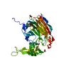
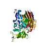
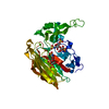
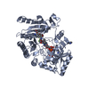
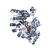
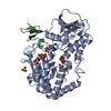
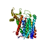
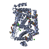
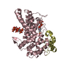
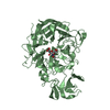
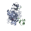
- PDB-3imn: Crystal structure of heparin lyase I from Bacteroides thetaiotaomicron -
+ Open data

- Basic information
| Entry | Database: PDB / ID: 3imn | ||||||
|---|---|---|---|---|---|---|---|
| Title | Crystal structure of heparin lyase I from Bacteroides thetaiotaomicron | ||||||
 Components Components | Heparin lyase I | ||||||
 Keywords Keywords | LYASE / jelly roll | ||||||
| Function / homology |  Function and homology information Function and homology information | ||||||
| Biological species |  Bacteroides thetaiotaomicron (bacteria) Bacteroides thetaiotaomicron (bacteria) | ||||||
| Method |  X-RAY DIFFRACTION / SYNCHROTRON / MAD / Resolution: 1.81 Å X-RAY DIFFRACTION / SYNCHROTRON / MAD / Resolution: 1.81 Å | ||||||
 Authors Authors | Han, Y.H. / Ryu, K.S. / Jeon, Y.H. | ||||||
 Citation Citation | Journal: J.Biol.Chem. / Year: 2009 Title: Structural snapshots of heparin depolymerization by heparin lyase I Authors: Han, Y.H. / Garron, M.L. / Kim, H.Y. / Kim, W.S. / Zhang, Z. / Ryu, K.S. / Shaya, D. / Xiao, Z. / Cheong, C. / Kim, Y.S. / Linhardt, R.J. / Jeon, Y.H. / Cygler, M. | ||||||
| History |
|
- Structure visualization
| Structure viewer | Molecule: MolmilJmol/JSmol |
|---|
- Downloads & links
-Download
| PDBx/mmCIF format | 3imn.cif.gz | 94.9 KB | Display | PDBx/mmCIF format |
|---|---|---|---|---|
| PDB format | pdb3imn.ent.gz | 70.9 KB | Display | PDB format |
| PDBx/mmJSON format | 3imn.json.gz | Tree view | PDBx/mmJSON format | |
| Others |  Other downloads Other downloads |
-Validation report
| Arichive directory | https://data.pdbj.org/pub/pdb/validation_reports/im/3imnftp://data.pdbj.org/pub/pdb/validation_reports/im/3imn | HTTPS FTP |
|---|
-Related structure data


-Links
 PDBj
PDBj- Assembly
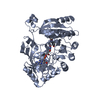
| Deposited unit | 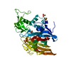
| ||||||||
|---|---|---|---|---|---|---|---|---|---|
| 1 |
| ||||||||
| Unit cell |
|
-Components
| #1: Protein | Mass: 42834.363 Da / Num. of mol.: 1 Source method: isolated from a genetically manipulated source Source: (gene. exp.) Bacteroides thetaiotaomicron (bacteria)Strain: WAL 2926 / Plasmid: pET22 / Production host: | ||||||
|---|---|---|---|---|---|---|---|
| #2: Chemical | ChemComp-SO4 /   Mass: 96.063 Da / Num. of mol.: 6 / Source method: obtained synthetically / Formula: SO4 Mass: 96.063 Da / Num. of mol.: 6 / Source method: obtained synthetically / Formula: SO4#3: Chemical | ChemComp-CA / |   Mass: 40.078 Da / Num. of mol.: 1 / Source method: obtained synthetically / Formula: Ca Mass: 40.078 Da / Num. of mol.: 1 / Source method: obtained synthetically / Formula: Ca#4: Water | ChemComp-HOH / |  Mass: 18.015 Da / Num. of mol.: 259 / Source method: isolated from a natural source / Formula: H2O Mass: 18.015 Da / Num. of mol.: 259 / Source method: isolated from a natural source / Formula: H2OSequence details | THERE IS NO UNP REFERENCE SEQUENCE DATABASE FOR THIS PROTEIN AT THE TIME OF PROCESSING. THE TWO ...THERE IS NO UNP REFERENCE SEQUENCE DATABASE FOR THIS PROTEIN AT THE TIME OF PROCESSING | |
-Experimental details
-Experiment
| Experiment | Method: X-RAY DIFFRACTION / Number of used crystals: 2 |
|---|
- Sample preparation
| Crystal | Density Matthews: 2.76 Å3/Da / Density % sol: 55.51 % / Mosaicity: 0.583 ° |
|---|---|
| Crystal grow | Temperature: 293 K / Method: vapor diffusion, hanging drop / pH: 5.5 Details: 0.1M Na citrate pH 5.5, 2.0M Ammonium sulfate, vapor diffusion, hanging drop, temperature 293K |
-Data collection
| Diffraction |
| |||||||||||||||||||||||||||||||||||||||||||||||||||||||||||||||||||||||||||||
|---|---|---|---|---|---|---|---|---|---|---|---|---|---|---|---|---|---|---|---|---|---|---|---|---|---|---|---|---|---|---|---|---|---|---|---|---|---|---|---|---|---|---|---|---|---|---|---|---|---|---|---|---|---|---|---|---|---|---|---|---|---|---|---|---|---|---|---|---|---|---|---|---|---|---|---|---|---|---|
| Diffraction source |
| |||||||||||||||||||||||||||||||||||||||||||||||||||||||||||||||||||||||||||||
| Detector |
| |||||||||||||||||||||||||||||||||||||||||||||||||||||||||||||||||||||||||||||
| Radiation |
| |||||||||||||||||||||||||||||||||||||||||||||||||||||||||||||||||||||||||||||
| Radiation wavelength |
| |||||||||||||||||||||||||||||||||||||||||||||||||||||||||||||||||||||||||||||
| Reflection | Redundancy: 6.4 % / Av σ(I) over netI: 37.14 / Number: 87493 / Rmerge(I) obs: 0.075 / Χ2: 1.04 / D res high: 2.5 Å / D res low: 50 Å / Num. obs: 13715 / % possible obs: 88.3 | |||||||||||||||||||||||||||||||||||||||||||||||||||||||||||||||||||||||||||||
| Diffraction reflection shell |
| |||||||||||||||||||||||||||||||||||||||||||||||||||||||||||||||||||||||||||||
| Reflection | Resolution: 1.8→58.93 Å / Num. all: 43830 / Num. obs: 40417 / % possible obs: 92.2 % / Redundancy: 4.4 % / Rmerge(I) obs: 0.057 / Χ2: 2.33 / Net I/av σ(I): 37.143 / Net I/σ(I): 21.3 / Num. measured all: 177991 | |||||||||||||||||||||||||||||||||||||||||||||||||||||||||||||||||||||||||||||
| Reflection shell |
|

- Processing
| Software |
| |||||||||||||||||||||||||||||||||||||||||||||||||||||||||||||||||
|---|---|---|---|---|---|---|---|---|---|---|---|---|---|---|---|---|---|---|---|---|---|---|---|---|---|---|---|---|---|---|---|---|---|---|---|---|---|---|---|---|---|---|---|---|---|---|---|---|---|---|---|---|---|---|---|---|---|---|---|---|---|---|---|---|---|---|
| Refinement | Method to determine structure: MAD / Resolution: 1.81→36.06 Å / Cor.coef. Fo:Fc: 0.953 / Cor.coef. Fo:Fc free: 0.938 / WRfactor Rfree: 0.213 / WRfactor Rwork: 0.179 / Occupancy max: 1 / Occupancy min: 1 / FOM work R set: 0.884 / SU B: 2.38 / SU ML: 0.074 / SU R Cruickshank DPI: 0.121 / SU Rfree: 0.118 / Cross valid method: THROUGHOUT / σ(F): 0 / ESU R: 0.121 / ESU R Free: 0.118 / Stereochemistry target values: MAXIMUM LIKELIHOOD Details: HYDROGENS HAVE BEEN ADDED IN THE RIDING POSITIONS U VALUES : REFINED INDIVIDUALLY
| |||||||||||||||||||||||||||||||||||||||||||||||||||||||||||||||||
| Solvent computation | Ion probe radii: 0.8 Å / Shrinkage radii: 0.8 Å / VDW probe radii: 1.4 Å / Solvent model: MASK | |||||||||||||||||||||||||||||||||||||||||||||||||||||||||||||||||
| Displacement parameters | Biso max: 80.48 Å2 / Biso mean: 27.651 Å2 / Biso min: 9.33 Å2
| |||||||||||||||||||||||||||||||||||||||||||||||||||||||||||||||||
| Refinement step | Cycle: LAST / Resolution: 1.81→36.06 Å
| |||||||||||||||||||||||||||||||||||||||||||||||||||||||||||||||||
| Refine LS restraints |
| |||||||||||||||||||||||||||||||||||||||||||||||||||||||||||||||||
| LS refinement shell | Resolution: 1.807→1.854 Å / Total num. of bins used: 20
|
