Type: MARMOSAIC 300 mm CCD / Detector: CCD / Date: Jun 20, 2009
Radiation
ID
Protocol
Monochromatic (M) / Laue (L)
Scattering type
Wavelength-ID
1
SINGLEWAVELENGTH
M
x-ray
1
2
SINGLEWAVELENGTH
M
x-ray
1
Radiation wavelength
ID
Wavelength (Å)
Relative weight
1
0.97946
1
2
1.07205
1
Reflection
Redundancy: 4.3 % / Av σ(I) over netI: 16.9 / Number: 58756 / Rmerge(I) obs: 0.093 / Χ2: 5.66 / D res high: 2 Å / D res low: 50 Å / Num. obs: 13539 / % possible obs: 98.9
Diffraction reflection shell
Highest resolution (Å)
Lowest resolution (Å)
% possible obs (%)
ID
Rmerge(I) obs
Chi squared
Redundancy
5.43
50
99.4
1
0.085
22.605
4.4
4.31
5.43
100
1
0.072
15.661
4.6
3.76
4.31
100
1
0.07
9.477
4.7
3.42
3.76
99.9
1
0.07
7.594
4.6
3.17
3.42
100
1
0.076
6.356
4.7
2.99
3.17
100
1
0.086
5.755
4.7
2.84
2.99
100
1
0.1
4.887
4.7
2.71
2.84
100
1
0.111
4.163
4.7
2.61
2.71
100
1
0.115
3.773
4.7
2.52
2.61
100
1
0.127
3.37
4.7
2.44
2.52
100
1
0.141
2.936
4.6
2.37
2.44
99.9
1
0.145
2.539
4.5
2.31
2.37
99.9
1
0.169
2.14
4.4
2.25
2.31
99.4
1
0.193
2.302
4.3
2.2
2.25
99.4
1
0.204
2.778
4.2
2.15
2.2
98.8
1
0.198
1.906
4
2.11
2.15
98.2
1
0.209
1.728
3.8
2.07
2.11
96.4
1
0.245
1.888
3.7
2.03
2.07
94.3
1
0.243
2.273
3.3
2
2.03
91.6
1
0.261
1.533
3.2
Reflection
Resolution: 1.5→50 Å / Num. obs: 31015 / % possible obs: 98.8 % / Redundancy: 5.7 % / Rmerge(I) obs: 0.042 / Χ2: 1.371 / Net I/σ(I): 17.3
Reflection shell
Resolution (Å)
Redundancy (%)
Rmerge(I) obs
Num. unique all
Χ2
Diffraction-ID
% possible all
1.5-1.53
2.5
0.239
1356
1.405
1,2
87.5
1.53-1.55
2.7
0.221
1481
1.117
1,2
93.4
1.55-1.58
3.1
0.213
1483
1.199
1,2
96.7
1.58-1.62
3.3
0.199
1554
1.258
1,2
98.6
1.62-1.65
3.6
0.196
1541
1.26
1,2
99.8
1.65-1.69
3.9
0.186
1548
1.432
1,2
99.7
1.69-1.73
4.1
0.186
1575
1.246
1,2
100
1.73-1.78
4.7
0.176
1540
1.247
1,2
100
1.78-1.83
5.4
0.172
1563
1.19
1,2
100
1.83-1.89
6.3
0.155
1576
1.206
1,2
100
1.89-1.96
7
0.124
1573
1.224
1,2
100
1.96-2.04
7.3
0.1
1554
1.288
1,2
100
2.04-2.13
7.5
0.086
1583
1.386
1,2
100
2.13-2.24
7.5
0.074
1569
1.5
1,2
100
2.24-2.38
7.5
0.061
1560
1.466
1,2
100
2.38-2.56
7.6
0.055
1570
1.484
1,2
100
2.56-2.82
7.6
0.047
1578
1.534
1,2
100
2.82-3.23
7.6
0.035
1587
1.495
1,2
100
3.23-4.07
7.5
0.028
1589
1.446
1,2
100
4.07-50
7.2
0.025
1635
1.422
1,2
99.9
-
Phasing
Phasing
Method: sad
-
Processing
Software
Name
Version
Classification
NB
DENZO
datareduction
SCALEPACK
datascaling
SHELX
phasing
REFMAC
refmac_5.5.0102
refinement
PDB_EXTRACT
3.005
dataextraction
HKL-2000
datareduction
HKL-2000
datascaling
Refinement
Method to determine structure: SAD / Resolution: 1.5→30 Å / Cor.coef. Fo:Fc: 0.954 / Cor.coef. Fo:Fc free: 0.931 / WRfactor Rfree: 0.266 / WRfactor Rwork: 0.211 / SU B: 1.548 / SU ML: 0.059 / Cross valid method: THROUGHOUT / σ(F): 0 / ESU R: 0.086 / ESU R Free: 0.094 / Stereochemistry target values: MAXIMUM LIKELIHOOD Details: A POTASSIUM HEXAIODOPLATIN-(IV)-ATE DERIVATIVE WAS USED FOR PHASING. HYDROGENS HAVE BEEN ADDED IN THE RIDING POSITIONS U VALUES: REFINED INDIVIDUALLY. ARP/WARP, COOT, MOLPROBITY WERE ALSO USED DURING REFINEMENT.
Rfactor
Num. reflection
% reflection
Selection details
Rfree
0.257
1038
3.4 %
THIN SHELLS (SFTOOLS)
Rwork
0.207
-
-
-
obs
0.209
30969
98.74 %
-
Solvent computation
Ion probe radii: 0.8 Å / Shrinkage radii: 0.8 Å / VDW probe radii: 1.4 Å / Solvent model: MASK
Displacement parameters
Biso mean: 15.078 Å2
Baniso -1
Baniso -2
Baniso -3
1-
0.24 Å2
0 Å2
-0.49 Å2
2-
-
-0.45 Å2
0 Å2
3-
-
-
0.12 Å2
Refinement step
Cycle: LAST / Resolution: 1.5→30 Å
Protein
Nucleic acid
Ligand
Solvent
Total
Num. atoms
1486
0
8
130
1624
Refine LS restraints
Refine-ID
Type
Dev ideal
Dev ideal target
Number
X-RAY DIFFRACTION
r_bond_refined_d
0.017
0.022
1609
X-RAY DIFFRACTION
r_bond_other_d
0.001
0.02
1046
X-RAY DIFFRACTION
r_angle_refined_deg
1.59
1.976
2198
X-RAY DIFFRACTION
r_angle_other_deg
0.874
3
2621
X-RAY DIFFRACTION
r_dihedral_angle_1_deg
5.457
5
219
X-RAY DIFFRACTION
r_dihedral_angle_2_deg
40.59
26.986
73
X-RAY DIFFRACTION
r_dihedral_angle_3_deg
12.894
15
318
X-RAY DIFFRACTION
r_dihedral_angle_4_deg
6.092
15
2
X-RAY DIFFRACTION
r_chiral_restr
0.086
0.2
269
X-RAY DIFFRACTION
r_gen_planes_refined
0.007
0.02
1789
X-RAY DIFFRACTION
r_gen_planes_other
0.002
0.02
289
X-RAY DIFFRACTION
r_mcbond_it
1.192
1.5
991
X-RAY DIFFRACTION
r_mcbond_other
0.309
1.5
400
X-RAY DIFFRACTION
r_mcangle_it
2.134
2
1623
X-RAY DIFFRACTION
r_scbond_it
3.128
3
618
X-RAY DIFFRACTION
r_scangle_it
5.052
4.5
561
LS refinement shell
Refine-ID: X-RAY DIFFRACTION / Total num. of bins used: 20
Resolution (Å)
Rfactor Rfree
Num. reflection Rfree
Rfactor Rwork
Num. reflection Rwork
Num. reflection all
% reflection obs (%)
1.5-1.54
0
0.305
2047
2301
88.961
1.54-1.582
0.294
102
0.272
2044
2239
95.846
1.582-1.628
0.313
89
0.246
2064
2178
98.852
1.628-1.678
0.253
97
0.24
2024
2128
99.671
1.678-1.732
0.284
87
0.219
1968
2058
99.854
1.732-1.793
0.257
61
0.215
1914
1979
99.798
1.793-1.861
0.256
64
0.202
1855
1921
99.896
1.861-1.936
0.23
20
0.205
1836
1859
99.839
1.936-2.022
0.253
83
0.193
1682
1768
99.83
2.022-2.12
0.234
69
0.193
1641
1710
100
2.12-2.234
0.271
48
0.195
1557
1605
100
2.234-2.369
0.19
44
0.186
1505
1549
100
2.369-2.531
0.214
36
0.197
1380
1416
100
2.531-2.732
0.218
42
0.194
1310
1352
100
2.732-2.991
0.316
52
0.222
1201
1253
100
2.991-3.34
0.245
36
0.215
1087
1123
100
3.34-3.848
0.265
18
0.196
985
1003
100
3.848-4.694
0.224
55
0.165
797
852
100
4.694-6.559
0.31
14
0.233
660
674
100
6.559-30
0.331
21
0.252
374
395
100
+
About Yorodumi
-
News
-
Feb 9, 2022. New format data for meta-information of EMDB entries
New format data for meta-information of EMDB entries
Version 3 of the EMDB header file is now the official format.
The previous official version 1.9 will be removed from the archive.
In the structure databanks used in Yorodumi, some data are registered as the other names, "COVID-19 virus" and "2019-nCoV". Here are the details of the virus and the list of structure data.
Jan 31, 2019. EMDB accession codes are about to change! (news from PDBe EMDB page)
EMDB accession codes are about to change! (news from PDBe EMDB page)
The allocation of 4 digits for EMDB accession codes will soon come to an end. Whilst these codes will remain in use, new EMDB accession codes will include an additional digit and will expand incrementally as the available range of codes is exhausted. The current 4-digit format prefixed with “EMD-” (i.e. EMD-XXXX) will advance to a 5-digit format (i.e. EMD-XXXXX), and so on. It is currently estimated that the 4-digit codes will be depleted around Spring 2019, at which point the 5-digit format will come into force.
The EM Navigator/Yorodumi systems omit the EMD- prefix.
Related info.:Q: What is EMD? / ID/Accession-code notation in Yorodumi/EM Navigator
Yorodumi is a browser for structure data from EMDB, PDB, SASBDB, etc.
This page is also the successor to EM Navigator detail page, and also detail information page/front-end page for Omokage search.
The word "yorodu" (or yorozu) is an old Japanese word meaning "ten thousand". "mi" (miru) is to see.
Related info.:EMDB / PDB / SASBDB / Comparison of 3 databanks / Yorodumi Search / Aug 31, 2016. New EM Navigator & Yorodumi / Yorodumi Papers / Jmol/JSmol / Function and homology information / Changes in new EM Navigator and Yorodumi
 Movie
Movie Controller

 Yorodumi
 Open data
 Basic information
 Components
 Keywords
 Function and homology information
 Homo sapiens (human)
 X-RAY DIFFRACTION /
  Authors
 Citation
 Structure visualization
 Downloads & links
 Other downloads
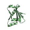


 PDBj
PDBj


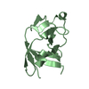
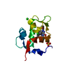
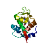
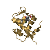
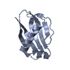
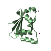
 Assembly
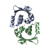
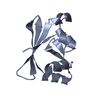


 Num. of mol.: 8 / Source method: obtained synthetically
 Mass: 18.015 Da / Num. of mol.: 130 / Source method: isolated from a natural source / Formula: H2O
 Sample preparation
 Processing