 Movie
Movie Controller

[English] 日本語
 Yorodumi
- PDB-1yn3: Crystal Structures of EAP Domains from Staphylococcus aureus Reve... -
+ Open data

- Basic information
| Entry | Database: PDB / ID: 1yn3 | ||||||
|---|---|---|---|---|---|---|---|
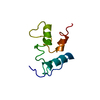
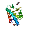
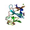
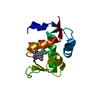
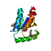
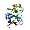
| Title | Crystal Structures of EAP Domains from Staphylococcus aureus Reveal an Unexpected Homology to Bacterial Superantigens | ||||||
 Components Components | truncated cell surface protein map-w | ||||||
 Keywords Keywords | TOXIN / PROTEIN BINDING / Virulence-factor / Extracellular-adherence protein / Staphylococcus | ||||||
| Function / homology | MAP domain / MAP domain / MAP repeat profile. / Ubiquitin-like (UB roll) - #120 / Ubiquitin-like (UB roll) / Roll / Alpha Beta / Protein map Function and homology information Function and homology information | ||||||
| Biological species |   Staphylococcus aureus (bacteria) Staphylococcus aureus (bacteria) | ||||||
| Method |  X-RAY DIFFRACTION / SYNCHROTRON / MOLECULAR REPLACEMENT / Resolution: 1.35 Å X-RAY DIFFRACTION / SYNCHROTRON / MOLECULAR REPLACEMENT / Resolution: 1.35 Å | ||||||
 Authors Authors | Geisbrecht, B.V. / Hamaoka, B.Y. / Perman, B. / Zemla, A. / Leahy, D.J. | ||||||
 Citation Citation | Journal: J.Biol.Chem. / Year: 2005 Title: The Crystal Structures of EAP Domains from Staphylococcus aureus Reveal an Unexpected Homology to Bacterial Superantigens. Authors: Geisbrecht, B.V. / Hamaoka, B.Y. / Perman, B. / Zemla, A. / Leahy, D.J. | ||||||
| History |
|
- Structure visualization
| Structure viewer | Molecule: MolmilJmol/JSmol |
|---|
- Downloads & links
-Download
| PDBx/mmCIF format | 1yn3.cif.gz | 56.3 KB | Display | PDBx/mmCIF format |
|---|---|---|---|---|
| PDB format | pdb1yn3.ent.gz | 41.5 KB | Display | PDB format |
| PDBx/mmJSON format | 1yn3.json.gz | Tree view | PDBx/mmJSON format | |
| Others |  Other downloads Other downloads |
-Validation report
| Arichive directory | https://data.pdbj.org/pub/pdb/validation_reports/yn/1yn3ftp://data.pdbj.org/pub/pdb/validation_reports/yn/1yn3 | HTTPS FTP |
|---|
-Related structure data
| Related structure data | 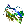 1yn4C 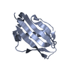 1yn5SC S: Starting model for refinement C: citing same article ( |
|---|---|
| Similar structure data |


-Links
 PDBj
PDBj- Assembly
| Deposited unit | 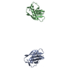
| ||||||||
|---|---|---|---|---|---|---|---|---|---|
| 1 | 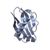
| ||||||||
| 2 | 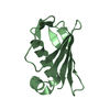
| ||||||||
| Unit cell |
|
-Components
| #1: Protein | Mass: 10810.184 Da / Num. of mol.: 2 / Fragment: residues 160-254 / Mutation: V253I Source method: isolated from a genetically manipulated source Source: (gene. exp.) Staphylococcus aureus (bacteria) / Strain: Mu50 / Plasmid: pET28 derivative / Production host: #2: Water | ChemComp-HOH / |  Mass: 18.015 Da / Num. of mol.: 304 / Source method: isolated from a natural source / Formula: H2O Mass: 18.015 Da / Num. of mol.: 304 / Source method: isolated from a natural source / Formula: H2O |
|---|
-Experimental details
-Experiment
| Experiment | Method: X-RAY DIFFRACTION / Number of used crystals: 1 |
|---|
- Sample preparation
| Crystal | Density Matthews: 2.59 Å3/Da / Density % sol: 52.56 % |
|---|---|
| Crystal grow | Temperature: 293 K / Method: vapor diffusion, hanging drop / pH: 6.4 Details: PEG 8000, ammonium sulfate, MES, pH 6.4, VAPOR DIFFUSION, HANGING DROP, temperature 293.0K |
-Data collection
| Diffraction | Mean temperature: 93 K |
|---|---|
| Diffraction source | Source: SYNCHROTRON / Site: NSLS  / Beamline: X4A / Wavelength: 0.9793 Å / Beamline: X4A / Wavelength: 0.9793 Å |
| Detector | Type: ADSC QUANTUM 4 / Detector: CCD |
| Radiation | Protocol: SINGLE WAVELENGTH / Monochromatic (M) / Laue (L): M / Scattering type: x-ray |
| Radiation wavelength | Wavelength: 0.9793 Å / Relative weight: 1 |
| Reflection | Resolution: 1.35→30 Å / Num. obs: 40720 / % possible obs: 88.8 % / Biso Wilson estimate: 13.9 Å2 |
| Reflection shell | Resolution: 1.35→1.45 Å / % possible all: 70.3 |
- Processing
| Software |
| ||||||||||||||||||||||||||||||||||||||||||||||||||||||||||||||||||||||||||||||||
|---|---|---|---|---|---|---|---|---|---|---|---|---|---|---|---|---|---|---|---|---|---|---|---|---|---|---|---|---|---|---|---|---|---|---|---|---|---|---|---|---|---|---|---|---|---|---|---|---|---|---|---|---|---|---|---|---|---|---|---|---|---|---|---|---|---|---|---|---|---|---|---|---|---|---|---|---|---|---|---|---|---|
| Refinement | Method to determine structure: MOLECULAR REPLACEMENT Starting model: EapH1, pdb entry 1YN5 Resolution: 1.35→29.87 Å / Rfactor Rfree error: 0.005 / Data cutoff high absF: 546804.14 / Data cutoff low absF: 0 / Isotropic thermal model: RESTRAINED / Cross valid method: THROUGHOUT / σ(F): 0 / Stereochemistry target values: Engh & Huber
| ||||||||||||||||||||||||||||||||||||||||||||||||||||||||||||||||||||||||||||||||
| Solvent computation | Solvent model: FLAT MODEL / Bsol: 46.4792 Å2 / ksol: 0.363177 e/Å3 | ||||||||||||||||||||||||||||||||||||||||||||||||||||||||||||||||||||||||||||||||
| Displacement parameters | Biso mean: 16.4 Å2
| ||||||||||||||||||||||||||||||||||||||||||||||||||||||||||||||||||||||||||||||||
| Refine analyze |
| ||||||||||||||||||||||||||||||||||||||||||||||||||||||||||||||||||||||||||||||||
| Refinement step | Cycle: LAST / Resolution: 1.35→29.87 Å
| ||||||||||||||||||||||||||||||||||||||||||||||||||||||||||||||||||||||||||||||||
| Refine LS restraints |
| ||||||||||||||||||||||||||||||||||||||||||||||||||||||||||||||||||||||||||||||||
| LS refinement shell | Resolution: 1.35→1.43 Å / Rfactor Rfree error: 0.018 / Total num. of bins used: 6
| ||||||||||||||||||||||||||||||||||||||||||||||||||||||||||||||||||||||||||||||||
| Xplor file |
|
