 Movie
Movie Controller

[English] 日本語
 Yorodumi
- PDB-2q3w: Ensemble refinement of the protein crystal structure of the cys84... -
+ Open data

- Basic information
| Entry | Database: PDB / ID: 2q3w | ||||||
|---|---|---|---|---|---|---|---|
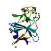
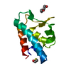
| Title | Ensemble refinement of the protein crystal structure of the cys84ala cys85ala double mutant of the [2Fe-2S] ferredoxin subunit of toluene-4-monooxygenase from Pseudomonas mendocina KR1 | ||||||
 Components Components | Toluene-4-monooxygenase system ferredoxin subunit | ||||||
 Keywords Keywords | ELECTRON TRANSPORT / Ensemble Refinement / Refinement Methodology Development / FERREDOXIN / FES / [2FE-2S] CLUSTER / RIESKE PROTEIN / TOLUENE-4-MONOOXYGENASE SUBUNIT / Structural Genomics / Protein Structure Initiative / PSI / Center for Eukaryotic Structural Genomics / CESG | ||||||
| Function / homology |  Function and homology information Function and homology informationtoluene catabolic process / 2 iron, 2 sulfur cluster binding / metal ion binding Similarity search - Function | ||||||
| Biological species |  Pseudomonas mendocina (bacteria) Pseudomonas mendocina (bacteria) | ||||||
| Method |  X-RAY DIFFRACTION / Re-refinement using ensemble model / Resolution: 1.48 Å X-RAY DIFFRACTION / Re-refinement using ensemble model / Resolution: 1.48 Å | ||||||
 Authors Authors | Levin, E.J. / Kondrashov, D.A. / Wesenberg, G.E. / Phillips Jr., G.N. / Center for Eukaryotic Structural Genomics (CESG) | ||||||
 Citation Citation | Journal: Structure / Year: 2007 Title: Ensemble refinement of protein crystal structures: validation and application. Authors: Levin, E.J. / Kondrashov, D.A. / Wesenberg, G.E. / Phillips, G.N. #1: Journal: ACTA CRYSTALLOGR.,SECT.D / Year: 2006Title: Structure of T4moC, the Rieske-type ferredoxin component of toluene 4-monooxygenase. Authors: Moe, L.A. / Bingman, C.A. / Wesenberg, G.E. / Phillips Jr., G.N. / Fox, B.G. | ||||||
| History |
|
- Structure visualization
| Structure viewer | Molecule: MolmilJmol/JSmol |
|---|
- Downloads & links
-Download
| PDBx/mmCIF format | 2q3w.cif.gz | 190.7 KB | Display | PDBx/mmCIF format |
|---|---|---|---|---|
| PDB format | pdb2q3w.ent.gz | 159.3 KB | Display | PDB format |
| PDBx/mmJSON format | 2q3w.json.gz | Tree view | PDBx/mmJSON format | |
| Others |  Other downloads Other downloads |
-Validation report
| Arichive directory | https://data.pdbj.org/pub/pdb/validation_reports/q3/2q3wftp://data.pdbj.org/pub/pdb/validation_reports/q3/2q3w | HTTPS FTP |
|---|
-Related structure data
| Related structure data | 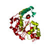 2q3mC  2q3oC 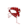 2q3pC  2q3qC 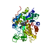 2q3rC 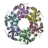 2q3sC 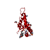 2q3tC 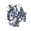 2q3uC 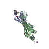 2q3vC  2q40C 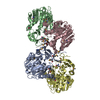 2q41C 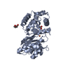 2q42C  2q43C 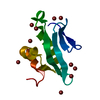 2q44C 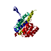 2q45C 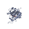 2q46C  2q47C  2q48C  2q49C  2q4aC  2q4bC  2q4cC 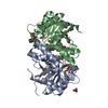 2q4dC  2q4eC  2q4fC  2q4hC 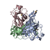 2q4iC  2q4jC 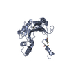 2q4kC  2q4lC 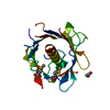 2q4mC  2q4nC  2q4oC 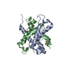 2q4pC  2q4qC  2q4rC  2q4sC  2q4tC  2q4uC 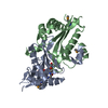 2q4vC 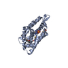 2q4xC  2q4yC 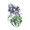 2q4zC 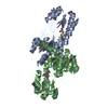 2q50C  2q51C 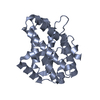 2q52C  1vm9S S: Starting model for refinement C: citing same article ( |
|---|---|
| Similar structure data | |
| Other databases |
-Links
 PDBj
PDBj


- Assembly
| Deposited unit | 
| ||||||||
|---|---|---|---|---|---|---|---|---|---|
| 1 |
| ||||||||
| Unit cell |
| ||||||||
| Number of models | 8 |
-Components
| #1: Protein | Mass: 12141.370 Da / Num. of mol.: 1 / Mutation: C84A,C85A Source method: isolated from a genetically manipulated source Source: (gene. exp.) Pseudomonas mendocina (bacteria) / Strain: KR1 / Gene: tmoC / Plasmid details: pET15b derivative / Plasmid: pT4CD15bTET(C84AC85A) / Species (production host): Escherichia coli / Production host: | ||||||
|---|---|---|---|---|---|---|---|
| #2: Chemical |   Mass: 24.305 Da / Num. of mol.: 2 / Source method: obtained synthetically / Formula: Mg Mass: 24.305 Da / Num. of mol.: 2 / Source method: obtained synthetically / Formula: Mg#3: Chemical | ChemComp-FES / |   Mass: 175.820 Da / Num. of mol.: 1 / Source method: obtained synthetically / Formula: Fe2S2 Mass: 175.820 Da / Num. of mol.: 1 / Source method: obtained synthetically / Formula: Fe2S2#4: Chemical |   Mass: 62.068 Da / Num. of mol.: 3 / Source method: obtained synthetically / Formula: C2H6O2 Mass: 62.068 Da / Num. of mol.: 3 / Source method: obtained synthetically / Formula: C2H6O2#5: Water | ChemComp-HOH / |  Mass: 18.015 Da / Num. of mol.: 133 / Source method: isolated from a natural source / Formula: H2O Mass: 18.015 Da / Num. of mol.: 133 / Source method: isolated from a natural source / Formula: H2O |
-Experimental details
-Experiment
| Experiment | Method: X-RAY DIFFRACTION |
|---|
- Sample preparation
| Crystal | Density Matthews: 2.01 Å3/Da / Density % sol: 38.67 % / Description: AUTHOR USED THE SF DATA FROM ENTRY 1VM9. |
|---|
-Data collection
| Radiation | Protocol: SINGLE WAVELENGTH / Monochromatic (M) / Laue (L): M / Scattering type: x-ray |
|---|---|
| Radiation wavelength | Relative weight: 1 |
- Processing
| Software |
| ||||||||||||||||||||||||||||||||||||||||||||||||||||||||||||||||||||||
|---|---|---|---|---|---|---|---|---|---|---|---|---|---|---|---|---|---|---|---|---|---|---|---|---|---|---|---|---|---|---|---|---|---|---|---|---|---|---|---|---|---|---|---|---|---|---|---|---|---|---|---|---|---|---|---|---|---|---|---|---|---|---|---|---|---|---|---|---|---|---|---|
| Refinement | Method to determine structure: Re-refinement using ensemble model Starting model: PDB entry 1VM9 Resolution: 1.48→31.43 Å / Rfactor Rfree error: 0.006 / Data cutoff high absF: 1803722.375 / Data cutoff low absF: 0 / Isotropic thermal model: RESTRAINED / Cross valid method: THROUGHOUT / σ(F): 0 Stereochemistry target values: maximum likelihood using amplitudes Details: This PDB entry is a re-refinement using an ensemble model of the previously deposited single-conformer structure 1vm9 and the first data set in the deposited structure factor file for 1vm9 ...Details: This PDB entry is a re-refinement using an ensemble model of the previously deposited single-conformer structure 1vm9 and the first data set in the deposited structure factor file for 1vm9 along with the R-free set defined therein. The coordinates were generated by an automated protocol from an initial model consisting of 8 identical copies of the protein and non-water hetero-atoms assigned fractional occupancies adding up to one, and a single copy of the solvent molecules. Refinement was carried out with all the conformers present simultaneously and with the potential energy terms corresponding to interactions between the different conformers excluded. The helix and sheet records were calculated using coordinates from the first conformer only. The structure visualization program PYMOL is well-suited for directly viewing the ensemble model presented in this PDB file.
| ||||||||||||||||||||||||||||||||||||||||||||||||||||||||||||||||||||||
| Solvent computation | Solvent model: FLAT MODEL / Bsol: 48.894 Å2 / ksol: 0.396 e/Å3 | ||||||||||||||||||||||||||||||||||||||||||||||||||||||||||||||||||||||
| Displacement parameters | Biso mean: 12.6 Å2
| ||||||||||||||||||||||||||||||||||||||||||||||||||||||||||||||||||||||
| Refine analyze |
| ||||||||||||||||||||||||||||||||||||||||||||||||||||||||||||||||||||||
| Refinement step | Cycle: LAST / Resolution: 1.48→31.43 Å
| ||||||||||||||||||||||||||||||||||||||||||||||||||||||||||||||||||||||
| Refine LS restraints |
| ||||||||||||||||||||||||||||||||||||||||||||||||||||||||||||||||||||||
| LS refinement shell | Refine-ID: X-RAY DIFFRACTION / Total num. of bins used: 6
| ||||||||||||||||||||||||||||||||||||||||||||||||||||||||||||||||||||||
| Xplor file |
|
