 Movie
Movie Controller


+ Open data

- Basic information
| Entry | Database: PDB / ID: 3i7k | ||||||
|---|---|---|---|---|---|---|---|
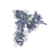
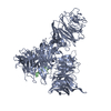
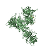
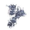
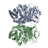
| Title | Crystal Structure of DDB1 in Complex with the H-Box Motif of WHX | ||||||
 Components Components |
| ||||||
 Keywords Keywords | PROTEIN BINDING/VIRAL PROTEIN / DDB1 / HBV / X protein / H-Box Motif / Cytoplasm / DNA damage / DNA repair / DNA-binding / Host-virus interaction / Nucleus / Phosphoprotein / Polymorphism / Ubl conjugation / Ubl conjugation pathway / Activator / Apoptosis / Mitochondrion / Transcription / Transcription regulation / PROTEIN BINDING-VIRAL PROTEIN COMPLEX | ||||||
| Function / homology |  Function and homology information Function and homology informationsymbiont-mediated activation of host NF-kappaB cascade / symbiont-mediated arrest of host cell cycle during G2/M transition / positive regulation by virus of viral protein levels in host cell / spindle assembly involved in female meiosis / epigenetic programming in the zygotic pronuclei / UV-damage excision repair / biological process involved in interaction with symbiont / regulation of mitotic cell cycle phase transition / WD40-repeat domain binding / Cul4A-RING E3 ubiquitin ligase complex ...symbiont-mediated activation of host NF-kappaB cascade / symbiont-mediated arrest of host cell cycle during G2/M transition / positive regulation by virus of viral protein levels in host cell / spindle assembly involved in female meiosis / epigenetic programming in the zygotic pronuclei / UV-damage excision repair / biological process involved in interaction with symbiont / regulation of mitotic cell cycle phase transition / WD40-repeat domain binding / Cul4A-RING E3 ubiquitin ligase complex / Cul4-RING E3 ubiquitin ligase complex / Cul4B-RING E3 ubiquitin ligase complex / ubiquitin ligase complex scaffold activity / negative regulation of reproductive process / negative regulation of developmental process / host cell mitochondrion / viral release from host cell / cullin family protein binding / ectopic germ cell programmed cell death / positive regulation of viral genome replication / positive regulation of gluconeogenesis / viral genome replication / proteasomal protein catabolic process / sperm end piece / nucleotide-excision repair / sperm principal piece / regulation of circadian rhythm / Recognition of DNA damage by PCNA-containing replication complex / DNA Damage Recognition in GG-NER / Wnt signaling pathway / Dual Incision in GG-NER / Transcription-Coupled Nucleotide Excision Repair (TC-NER) / Formation of TC-NER Pre-Incision Complex / Formation of Incision Complex in GG-NER / positive regulation of protein catabolic process / cellular response to UV / Dual incision in TC-NER / Gap-filling DNA repair synthesis and ligation in TC-NER / rhythmic process / sperm midpiece / site of double-strand break / Neddylation / ubiquitin-dependent protein catabolic process / damaged DNA binding / proteasome-mediated ubiquitin-dependent protein catabolic process / protein-macromolecule adaptor activity / chromosome, telomeric region / protein ubiquitination / DNA repair / apoptotic process / DNA damage response / negative regulation of apoptotic process / nucleolus / host cell nucleus / protein-containing complex binding / DNA-templated transcription / protein-containing complex / : / DNA binding / extracellular exosome / nucleoplasm / nucleus / cytoplasm Similarity search - Function | ||||||
| Biological species |  Homo sapiens (human) Homo sapiens (human) Woodchuck hepatitis B virus Woodchuck hepatitis B virus | ||||||
| Method |  X-RAY DIFFRACTION / SYNCHROTRON / MOLECULAR REPLACEMENT / Resolution: 2.8 Å X-RAY DIFFRACTION / SYNCHROTRON / MOLECULAR REPLACEMENT / Resolution: 2.8 Å | ||||||
 Authors Authors | Li, T. / Robert, E.I. / Breugel, P.C.V. / Strubin, M. / Zheng, N. | ||||||
 Citation Citation | Journal: Nat.Struct.Mol.Biol. / Year: 2010 Title: A promiscuous alpha-helical motif anchors viral hijackers and substrate receptors to the CUL4-DDB1 ubiquitin ligase machinery. Authors: Li, T. / Robert, E.I. / van Breugel, P.C. / Strubin, M. / Zheng, N. | ||||||
| History |
|
- Structure visualization
| Structure viewer | Molecule: MolmilJmol/JSmol |
|---|
- Downloads & links
-Download
| PDBx/mmCIF format | 3i7k.cif.gz | 215.5 KB | Display | PDBx/mmCIF format |
|---|---|---|---|---|
| PDB format | pdb3i7k.ent.gz | 170.6 KB | Display | PDB format |
| PDBx/mmJSON format | 3i7k.json.gz | Tree view | PDBx/mmJSON format | |
| Others |  Other downloads Other downloads |
-Validation report
| Arichive directory | https://data.pdbj.org/pub/pdb/validation_reports/i7/3i7kftp://data.pdbj.org/pub/pdb/validation_reports/i7/3i7k | HTTPS FTP |
|---|
-Related structure data
| Related structure data |  3i7hC 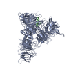 3i7lC 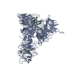 3i7nC 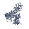 3i7oC  3i7pC 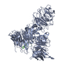 3i89C 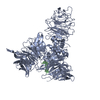 3i8cC 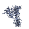 3i8eC 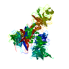 2b5mS S: Starting model for refinement C: citing same article ( |
|---|---|
| Similar structure data |


-Links
 PDBj
PDBj


- Assembly
| Deposited unit | 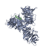
| ||||||||
|---|---|---|---|---|---|---|---|---|---|
| 1 |
| ||||||||
| Unit cell |
|
-Components
| #1: Protein | Mass: 127399.766 Da / Num. of mol.: 1 Source method: isolated from a genetically manipulated source Source: (gene. exp.) Homo sapiens (human) / Gene: DDB1, XAP1 / Production host:   Spodoptera frugiperda (fall armyworm) / References: UniProt: Q16531 Spodoptera frugiperda (fall armyworm) / References: UniProt: Q16531 |
|---|---|
| #2: Protein/peptide | Mass: 1658.880 Da / Num. of mol.: 1 / Fragment: Residues 86-99 / Source method: obtained synthetically / Source: (synth.) Woodchuck hepatitis B virus / References: UniProt: Q89246, UniProt: P03167*PLUS |
| Has protein modification | Y |
| Sequence details | MUTATIONS OCCURRED DURING THE CLONING OF DDB1 |
-Experimental details
-Experiment
| Experiment | Method: X-RAY DIFFRACTION / Number of used crystals: 1 |
|---|
- Sample preparation
| Crystal | Density Matthews: 3.02 Å3/Da / Density % sol: 59.27 % |
|---|---|
| Crystal grow | Temperature: 277 K / Method: vapor diffusion, hanging drop / pH: 6.5 Details: 16% PEG 4000, 0.2M SODIUM CHLORIDE, 0.1M MES, 0.005 M DTT, pH 6.5, VAPOR DIFFUSION, HANGING DROP, temperature 277K |
-Data collection
| Diffraction | Mean temperature: 173 K | |||||||||
|---|---|---|---|---|---|---|---|---|---|---|
| Diffraction source | Source: SYNCHROTRON / Site: ALS  / Beamline: 5.0.2 / Wavelength: 1.0,1.005 / Beamline: 5.0.2 / Wavelength: 1.0,1.005 | |||||||||
| Detector | Type: ADSC QUANTUM 315 / Detector: CCD / Date: May 28, 2008 | |||||||||
| Radiation | Protocol: SINGLE WAVELENGTH / Monochromatic (M) / Laue (L): M / Scattering type: x-ray | |||||||||
| Radiation wavelength |
| |||||||||
| Reflection | Resolution: 2.8→46.03 Å / Num. all: 38728 / Num. obs: 35137 / % possible obs: 90.7 % / Observed criterion σ(F): 2 / Observed criterion σ(I): 1 / Redundancy: 4 % / Rmerge(I) obs: 0.07 / Net I/σ(I): 23.4 | |||||||||
| Reflection shell | Resolution: 2.8→2.87 Å / Rmerge(I) obs: 0.426 / Mean I/σ(I) obs: 2.6 / Num. unique all: 2285 / % possible all: 94.6 |
- Processing
| Software |
| ||||||||||||||||||||||||||||||||||||||||||||||||||||||||||||||||||||||
|---|---|---|---|---|---|---|---|---|---|---|---|---|---|---|---|---|---|---|---|---|---|---|---|---|---|---|---|---|---|---|---|---|---|---|---|---|---|---|---|---|---|---|---|---|---|---|---|---|---|---|---|---|---|---|---|---|---|---|---|---|---|---|---|---|---|---|---|---|---|---|---|
| Refinement | Method to determine structure: MOLECULAR REPLACEMENT Starting model: PDB ENTRY 2B5M Resolution: 2.8→46.03 Å / Cor.coef. Fo:Fc: 0.916 / Cor.coef. Fo:Fc free: 0.888 / SU B: 14.606 / SU ML: 0.289 / Cross valid method: THROUGHOUT / σ(F): 2 / ESU R: 0.679 / ESU R Free: 0.396 / Stereochemistry target values: MAXIMUM LIKELIHOOD / Details: HYDROGENS HAVE BEEN ADDED IN THE RIDING POSITIONS
| ||||||||||||||||||||||||||||||||||||||||||||||||||||||||||||||||||||||
| Solvent computation | Ion probe radii: 0.8 Å / Shrinkage radii: 0.8 Å / VDW probe radii: 1.2 Å / Solvent model: MASK | ||||||||||||||||||||||||||||||||||||||||||||||||||||||||||||||||||||||
| Displacement parameters | Biso mean: 71.954 Å2
| ||||||||||||||||||||||||||||||||||||||||||||||||||||||||||||||||||||||
| Refinement step | Cycle: LAST / Resolution: 2.8→46.03 Å
| ||||||||||||||||||||||||||||||||||||||||||||||||||||||||||||||||||||||
| Refine LS restraints |
| ||||||||||||||||||||||||||||||||||||||||||||||||||||||||||||||||||||||
| LS refinement shell | Resolution: 2.8→2.872 Å / Total num. of bins used: 20
|
