 Movie
Movie Controller


+ Open data

- Basic information
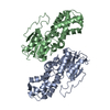
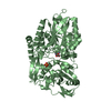
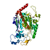
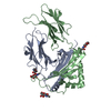
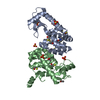
| Entry | Database: PDB / ID: 3hzt | ||||||
|---|---|---|---|---|---|---|---|
| Title | Crystal structure of Toxoplasma gondii CDPK3, TGME49_105860 | ||||||
 Components Components | Calcium-dependent protein kinase 3 | ||||||
 Keywords Keywords | TRANSFERASE / calcium dependent kinase / calmodulin / troponin parasite / Structural Genomics / Structural Genomics Consortium / SGC / ATP-binding / Kinase / Nucleotide-binding / Serine/threonine-protein kinase | ||||||
| Function / homology |  Function and homology information Function and homology informationsymbiont-containing vacuole membrane / motile cilium / non-specific serine/threonine protein kinase / protein serine/threonine kinase activity / calcium ion binding / host cell plasma membrane / ATP binding / plasma membrane Similarity search - Function | ||||||
| Biological species |  | ||||||
| Method |  X-RAY DIFFRACTION / SYNCHROTRON / MOLECULAR REPLACEMENT / molecular replacement / Resolution: 2 Å X-RAY DIFFRACTION / SYNCHROTRON / MOLECULAR REPLACEMENT / molecular replacement / Resolution: 2 Å | ||||||
 Authors Authors | Wernimont, A.K. / Artz, J.D. / Finnerty, P. / Wasney, G. / Allali-Hassani, A. / Vedadi, M. / Bochkarev, A. / Arrowsmith, C.H. / Edwards, A.M. / Bountra, C. ...Wernimont, A.K. / Artz, J.D. / Finnerty, P. / Wasney, G. / Allali-Hassani, A. / Vedadi, M. / Bochkarev, A. / Arrowsmith, C.H. / Edwards, A.M. / Bountra, C. / Weigelt, J. / Hui, R. / Amani, M. / Structural Genomics Consortium (SGC) | ||||||
 Citation Citation | Journal: Nat.Struct.Mol.Biol. / Year: 2010 Title: Structures of apicomplexan calcium-dependent protein kinases reveal mechanism of activation by calcium. Authors: Wernimont, A.K. / Artz, J.D. / Finerty, P. / Lin, Y.H. / Amani, M. / Allali-Hassani, A. / Senisterra, G. / Vedadi, M. / Tempel, W. / Mackenzie, F. / Chau, I. / Lourido, S. / Sibley, L.D. / Hui, R. | ||||||
| History |
|
- Structure visualization
| Structure viewer | Molecule: MolmilJmol/JSmol |
|---|
- Downloads & links
-Download
| PDBx/mmCIF format | 3hzt.cif.gz | 108.3 KB | Display | PDBx/mmCIF format |
|---|---|---|---|---|
| PDB format | pdb3hzt.ent.gz | 79.4 KB | Display | PDB format |
| PDBx/mmJSON format | 3hzt.json.gz | Tree view | PDBx/mmJSON format | |
| Others |  Other downloads Other downloads |
-Validation report
| Arichive directory | https://data.pdbj.org/pub/pdb/validation_reports/hz/3hztftp://data.pdbj.org/pub/pdb/validation_reports/hz/3hzt | HTTPS FTP |
|---|
-Related structure data
| Related structure data | 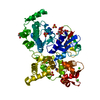 3hx4C 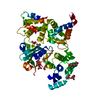 3igoC 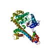 3ku2C 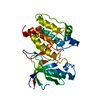 3dxnS C: citing same article ( S: Starting model for refinement |
|---|---|
| Similar structure data |


-Links
 PDBj
PDBj
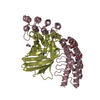
- Assembly
| Deposited unit | 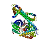
| ||||||||
|---|---|---|---|---|---|---|---|---|---|
| 1 |
| ||||||||
| Unit cell |
|
-Components
| #1: Protein | Mass: 53426.926 Da / Num. of mol.: 1 / Fragment: UNP residues 72-537 Source method: isolated from a genetically manipulated source Source: (gene. exp.)  References: UniProt: B6KR85, UniProt: Q3HNM6*PLUS, Ca2+/calmodulin-dependent protein kinase | ||||
|---|---|---|---|---|---|
| #2: Chemical | ChemComp-MG /   Mass: 24.305 Da / Num. of mol.: 1 / Source method: obtained synthetically / Formula: Mg Mass: 24.305 Da / Num. of mol.: 1 / Source method: obtained synthetically / Formula: Mg | ||||
| #3: Chemical | ChemComp-GOL /   Mass: 92.094 Da / Num. of mol.: 8 / Source method: obtained synthetically / Formula: C3H8O3 Mass: 92.094 Da / Num. of mol.: 8 / Source method: obtained synthetically / Formula: C3H8O3#4: Chemical | ChemComp-J60 / | 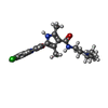  Mass: 414.928 Da / Num. of mol.: 1 / Source method: obtained synthetically / Formula: C22H27ClN4O2 Mass: 414.928 Da / Num. of mol.: 1 / Source method: obtained synthetically / Formula: C22H27ClN4O2#5: Water | ChemComp-HOH / |  Mass: 18.015 Da / Num. of mol.: 202 / Source method: isolated from a natural source / Formula: H2O Mass: 18.015 Da / Num. of mol.: 202 / Source method: isolated from a natural source / Formula: H2O |
-Experimental details
-Experiment
| Experiment | Method: X-RAY DIFFRACTION / Number of used crystals: 1 |
|---|
- Sample preparation
| Crystal | Density Matthews: 2.45 Å3/Da / Density % sol: 49.8 % |
|---|---|
| Crystal grow | Temperature: 293 K / Method: vapor diffusion, hanging drop / pH: 7.5 Details: 18% PEG 3350, 0.2 M KF, 3 mM SU11652, Glycerol, pH 7.5, VAPOR DIFFUSION, HANGING DROP, temperature 293K |
-Data collection
| Diffraction | Mean temperature: 100 K |
|---|---|
| Diffraction source | Source: SYNCHROTRON / Site: APS  / Beamline: 19-ID / Wavelength: 0.97937 Å / Beamline: 19-ID / Wavelength: 0.97937 Å |
| Detector | Type: ADSC QUANTUM 315 / Detector: CCD / Date: Feb 15, 2009 |
| Radiation | Protocol: SINGLE WAVELENGTH / Monochromatic (M) / Laue (L): M / Scattering type: x-ray |
| Radiation wavelength | Wavelength: 0.97937 Å / Relative weight: 1 |
| Reflection | Resolution: 2→30 Å / Num. all: 35409 / Num. obs: 35409 / % possible obs: 99.7 % / Observed criterion σ(F): 0 / Observed criterion σ(I): 0 / Redundancy: 3.6 % / Biso Wilson estimate: 26.9 Å2 / Rmerge(I) obs: 0.048 / Rsym value: 0.033 / Χ2: 1.12 / Net I/σ(I): 26.63 |
| Reflection shell | Resolution: 2→2.03 Å / Redundancy: 3.3 % / Rmerge(I) obs: 0.457 / Mean I/σ(I) obs: 2.686 / Num. unique all: 1693 / Rsym value: 0.369 / Χ2: 1.273 / % possible all: 96.6 |
-Phasing
| Phasing | Method: molecular replacement |
|---|
- Processing
| Software |
| |||||||||||||||||||||||||||||||||||||||||||||||||||||||||||||||||
|---|---|---|---|---|---|---|---|---|---|---|---|---|---|---|---|---|---|---|---|---|---|---|---|---|---|---|---|---|---|---|---|---|---|---|---|---|---|---|---|---|---|---|---|---|---|---|---|---|---|---|---|---|---|---|---|---|---|---|---|---|---|---|---|---|---|---|
| Refinement | Method to determine structure: MOLECULAR REPLACEMENT Starting model: PDB entry 3DXN Resolution: 2→30 Å / Cor.coef. Fo:Fc: 0.949 / Cor.coef. Fo:Fc free: 0.922 / WRfactor Rfree: 0.257 / WRfactor Rwork: 0.207 / Occupancy max: 1 / Occupancy min: 0.2 / FOM work R set: 0.829 / SU B: 4.157 / SU ML: 0.118 / SU R Cruickshank DPI: 0.185 / SU Rfree: 0.169 / Cross valid method: THROUGHOUT / σ(F): 0 / ESU R: 0.185 / ESU R Free: 0.169 / Stereochemistry target values: MAXIMUM LIKELIHOOD Details: 1. HYDROGENS HAVE BEEN ADDED IN THE RIDING POSITIONS. 2. U VALUES: REFINED INDIVIDUALLY.
| |||||||||||||||||||||||||||||||||||||||||||||||||||||||||||||||||
| Solvent computation | Ion probe radii: 0.8 Å / Shrinkage radii: 0.8 Å / VDW probe radii: 1.2 Å / Solvent model: MASK | |||||||||||||||||||||||||||||||||||||||||||||||||||||||||||||||||
| Displacement parameters | Biso max: 93.18 Å2 / Biso mean: 42.411 Å2 / Biso min: 13.87 Å2
| |||||||||||||||||||||||||||||||||||||||||||||||||||||||||||||||||
| Refinement step | Cycle: LAST / Resolution: 2→30 Å
| |||||||||||||||||||||||||||||||||||||||||||||||||||||||||||||||||
| Refine LS restraints |
| |||||||||||||||||||||||||||||||||||||||||||||||||||||||||||||||||
| LS refinement shell | Resolution: 2→2.05 Å / Total num. of bins used: 20
|
