 Movie
Movie Controller

[English] 日本語
 Yorodumi
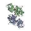
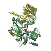
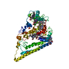
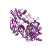
- PDB-3hv1: Crystal structure of a polar amino acid ABC uptake transporter su... -
+ Open data

- Basic information
| Entry | Database: PDB / ID: 3hv1 | ||||||
|---|---|---|---|---|---|---|---|
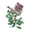
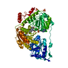
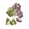
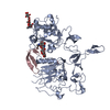
| Title | Crystal structure of a polar amino acid ABC uptake transporter substrate binding protein from Streptococcus thermophilus | ||||||
 Components Components | Polar amino acid ABC uptake transporter substrate binding protein | ||||||
 Keywords Keywords | TRANSPORT PROTEIN / Polar amino acid / ABC uptake transporter / Protein Structure Initiative II(PSI II) / NYSGXRC / 11316l / Structural Genomics / New York SGX Research Center for Structural Genomics | ||||||
| Function / homology | Bacterial periplasmic substrate-binding proteins / Bacterial extracellular solute-binding proteins, family 3 / Solute-binding protein family 3/N-terminal domain of MltF / Periplasmic binding protein-like II / D-Maltodextrin-Binding Protein; domain 2 / Prokaryotic membrane lipoprotein lipid attachment site profile. / 3-Layer(aba) Sandwich / Alpha Beta / Polar amino acid ABC uptake transporter substrate binding protein Function and homology information Function and homology information | ||||||
| Biological species |  Streptococcus thermophilus LMG 18311 (bacteria) Streptococcus thermophilus LMG 18311 (bacteria) | ||||||
| Method |  X-RAY DIFFRACTION / SYNCHROTRON / SAD / Resolution: 1.9 Å X-RAY DIFFRACTION / SYNCHROTRON / SAD / Resolution: 1.9 Å | ||||||
 Authors Authors | Palani, K. / Burley, S.K. / Swaminathan, S. / New York SGX Research Center for Structural Genomics (NYSGXRC) | ||||||
 Citation Citation | Journal: To be Published Title: Crystal structure of a polar amino acid ABC uptake transporter substrate binding protein from Streptococcus thermophilus Authors: Palani, K. / Burley, S.K. / Swaminathan, S. | ||||||
| History |
|
- Structure visualization
| Structure viewer | Molecule: MolmilJmol/JSmol |
|---|
- Downloads & links
-Download
| PDBx/mmCIF format | 3hv1.cif.gz | 120.9 KB | Display | PDBx/mmCIF format |
|---|---|---|---|---|
| PDB format | pdb3hv1.ent.gz | 93.3 KB | Display | PDB format |
| PDBx/mmJSON format | 3hv1.json.gz | Tree view | PDBx/mmJSON format | |
| Others |  Other downloads Other downloads |
-Validation report
| Summary document | 3hv1_validation.pdf.gz | 433.4 KB | Display | wwPDB validaton report |
|---|---|---|---|---|
| Full document | 3hv1_full_validation.pdf.gz | 441.3 KB | Display | |
| Data in XML | 3hv1_validation.xml.gz | 23.4 KB | Display | |
| Data in CIF | 3hv1_validation.cif.gz | 33.5 KB | Display | |
| Arichive directory | https://data.pdbj.org/pub/pdb/validation_reports/hv/3hv1ftp://data.pdbj.org/pub/pdb/validation_reports/hv/3hv1 | HTTPS FTP |
-Related structure data
| Similar structure data | |
|---|---|
| Other databases |


-Links
 PDBj
PDBj


- Assembly
| Deposited unit | 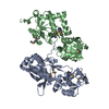
| ||||||||
|---|---|---|---|---|---|---|---|---|---|
| 1 |
| ||||||||
| 2 | 
| ||||||||
| 3 | 
| ||||||||
| Unit cell |
|
-Components
| #1: Protein | Mass: 30505.230 Da / Num. of mol.: 2 / Fragment: residues 27-283 Source method: isolated from a genetically manipulated source Source: (gene. exp.) Streptococcus thermophilus LMG 18311 (bacteria)Gene: stu0877 / Plasmid: BC-pSGX3 (BC) / Production host: #2: Water | ChemComp-HOH / |  Mass: 18.015 Da / Num. of mol.: 281 / Source method: isolated from a natural source / Formula: H2O Mass: 18.015 Da / Num. of mol.: 281 / Source method: isolated from a natural source / Formula: H2OHas protein modification | Y | |
|---|
-Experimental details
-Experiment
| Experiment | Method: X-RAY DIFFRACTION / Number of used crystals: 1 |
|---|
- Sample preparation
| Crystal | Density Matthews: 2.36 Å3/Da / Density % sol: 47.94 % |
|---|---|
| Crystal grow | Temperature: 298 K / Method: vapor diffusion, sitting drop / pH: 7.5 Details: 0.2M L-Proline, 0.1M HEPES, 10% Polyethylene glycol monomethyl ether 2000, pH 7.5, VAPOR DIFFUSION, SITTING DROP, temperature 298.0K |
-Data collection
| Diffraction | Mean temperature: 100 K |
|---|---|
| Diffraction source | Source: SYNCHROTRON / Site: NSLS  / Beamline: X29A / Wavelength: 0.979 Å / Beamline: X29A / Wavelength: 0.979 Å |
| Detector | Type: ADSC QUANTUM 315 / Detector: CCD / Date: Jun 4, 2009 / Details: MIRRORS |
| Radiation | Monochromator: Si(III) Channel / Protocol: SINGLE WAVELENGTH / Monochromatic (M) / Laue (L): M / Scattering type: x-ray |
| Radiation wavelength | Wavelength: 0.979 Å / Relative weight: 1 |
| Reflection | Resolution: 1.9→45.02 Å / Num. all: 44250 / Num. obs: 44250 / % possible obs: 98.7 % / Observed criterion σ(F): 0 / Observed criterion σ(I): 0 / Redundancy: 7.4 % / Biso Wilson estimate: 18.9 Å2 / Rmerge(I) obs: 0.055 / Net I/σ(I): 19.5 |
| Reflection shell | Resolution: 1.9→1.97 Å / Redundancy: 7.4 % / Rmerge(I) obs: 0.378 / Mean I/σ(I) obs: 2.5 / Num. unique all: 4333 / % possible all: 97.9 |
- Processing
| Software |
| |||||||||||||||||||||||||
|---|---|---|---|---|---|---|---|---|---|---|---|---|---|---|---|---|---|---|---|---|---|---|---|---|---|---|
| Refinement | Method to determine structure: SAD / Resolution: 1.9→45.02 Å / Rfactor Rfree error: 0.005 / Data cutoff high absF: 248640.19 / Data cutoff low absF: 0 / Isotropic thermal model: RESTRAINED / Cross valid method: THROUGHOUT / σ(F): 0 / Stereochemistry target values: Engh & Huber
| |||||||||||||||||||||||||
| Solvent computation | Solvent model: FLAT MODEL / Bsol: 43.0617 Å2 / ksol: 0.353258 e/Å3 | |||||||||||||||||||||||||
| Displacement parameters | Biso mean: 32.6 Å2
| |||||||||||||||||||||||||
| Refine analyze |
| |||||||||||||||||||||||||
| Refinement step | Cycle: LAST / Resolution: 1.9→45.02 Å
| |||||||||||||||||||||||||
| Refine LS restraints |
| |||||||||||||||||||||||||
| LS refinement shell | Resolution: 1.9→2.02 Å / Rfactor Rfree error: 0.018 / Total num. of bins used: 6
| |||||||||||||||||||||||||
| Xplor file |
|
