 Movie
Movie Controller

[English] 日本語
 Yorodumi
- PDB-3hki: Crystal structure of murine thrombin mutant W215A/E217A in comple... -
+ Open data

- Basic information
| Entry | Database: PDB / ID: 3hki | ||||||
|---|---|---|---|---|---|---|---|
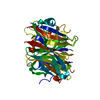
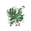
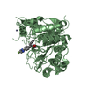
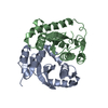
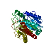
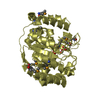
| Title | Crystal structure of murine thrombin mutant W215A/E217A in complex with the extracellular fragment of human PAR1 | ||||||
 Components Components |
| ||||||
 Keywords Keywords | HYDROLASE / Serine protease / Acute phase / Blood coagulation / Cleavage on pair of basic residues / Disulfide bond / Gamma-carboxyglutamic acid / Glycoprotein / Kringle / Protease / Zymogen / Cell membrane / G-protein coupled receptor / Membrane / Phosphoprotein / Receptor / Transducer / Transmembrane | ||||||
| Function / homology |  Function and homology information Function and homology information: / Platelet Aggregation (Plug Formation) / Gamma-carboxylation of protein precursors / Transport of gamma-carboxylated protein precursors from the endoplasmic reticulum to the Golgi apparatus / : / Removal of aminoterminal propeptides from gamma-carboxylated proteins / negative regulation of renin secretion into blood stream / dendritic cell homeostasis / trans-synaptic signaling by endocannabinoid, modulating synaptic transmission / platelet dense tubular network ...: / Platelet Aggregation (Plug Formation) / Gamma-carboxylation of protein precursors / Transport of gamma-carboxylated protein precursors from the endoplasmic reticulum to the Golgi apparatus / : / Removal of aminoterminal propeptides from gamma-carboxylated proteins / negative regulation of renin secretion into blood stream / dendritic cell homeostasis / trans-synaptic signaling by endocannabinoid, modulating synaptic transmission / platelet dense tubular network / establishment of synaptic specificity at neuromuscular junction / thrombin-activated receptor activity / Thrombin signalling through proteinase activated receptors (PARs) / Regulation of Complement cascade / connective tissue replacement involved in inflammatory response wound healing / regulation of interleukin-1 beta production / platelet dense granule organization / Peptide ligand-binding receptors / G alpha (q) signalling events / Cell surface interactions at the vascular wall / cell-cell junction maintenance / positive regulation of smooth muscle contraction / positive regulation of calcium ion transport / : / thrombospondin receptor activity / thrombin / thrombin-activated receptor signaling pathway / negative regulation of glomerular filtration / negative regulation of astrocyte differentiation / regulation of blood coagulation / neutrophil-mediated killing of gram-negative bacterium / positive regulation of phospholipase C-activating G protein-coupled receptor signaling pathway / positive regulation of Rho protein signal transduction / positive regulation of collagen biosynthetic process / negative regulation of blood coagulation / positive regulation of blood coagulation / positive regulation of vasoconstriction / anatomical structure morphogenesis / G-protein alpha-subunit binding / : / regulation of cytosolic calcium ion concentration / fibrinolysis / negative regulation of proteolysis / release of sequestered calcium ion into cytosol / negative regulation of cytokine production involved in inflammatory response / homeostasis of number of cells within a tissue / acute-phase response / Peptide ligand-binding receptors / positive regulation of release of sequestered calcium ion into cytosol / positive regulation of interleukin-8 production / positive regulation of receptor signaling pathway via JAK-STAT / neuromuscular junction / lipopolysaccharide binding / platelet activation / caveola / positive regulation of protein localization to nucleus / response to wounding / regulation of synaptic plasticity / positive regulation of interleukin-6 production / G protein-coupled receptor activity / positive regulation of reactive oxygen species metabolic process / late endosome / peptidase activity / regulation of cell shape / antimicrobial humoral immune response mediated by antimicrobial peptide / heparin binding / G-protein beta-subunit binding / Thrombin signalling through proteinase activated receptors (PARs) / extracellular matrix / positive regulation of cytosolic calcium ion concentration / positive regulation of cell growth / regulation of gene expression / endopeptidase activity / response to lipopolysaccharide / phospholipase C-activating G protein-coupled receptor signaling pathway / G alpha (q) signalling events / negative regulation of neuron apoptotic process / early endosome / positive regulation of ERK1 and ERK2 cascade / positive regulation of canonical NF-kappaB signal transduction / positive regulation of MAPK cascade / postsynaptic membrane / cell surface receptor signaling pathway / positive regulation of phosphatidylinositol 3-kinase/protein kinase B signal transduction / positive regulation of cell migration / positive regulation of apoptotic process / G protein-coupled receptor signaling pathway / inflammatory response / receptor ligand activity / serine-type endopeptidase activity / signaling receptor binding / external side of plasma membrane / negative regulation of cell population proliferation / calcium ion binding / positive regulation of cell population proliferation / positive regulation of DNA-templated transcription / cell surface / Golgi apparatus / proteolysis / : Similarity search - Function | ||||||
| Biological species |   Homo sapiens (human) Homo sapiens (human) | ||||||
| Method |  X-RAY DIFFRACTION / SYNCHROTRON / MOLECULAR REPLACEMENT / Resolution: 2.2 Å X-RAY DIFFRACTION / SYNCHROTRON / MOLECULAR REPLACEMENT / Resolution: 2.2 Å | ||||||
 Authors Authors | Gandhi, P.S. / Page, M.J. / Chen, Z. / Bush-Pelc, L. / Di Cera, E. | ||||||
 Citation Citation | Journal: J.Biol.Chem. / Year: 2009 Title: Mechanism of the Anticoagulant Activity of Thrombin Mutant W215A/E217A. Authors: Gandhi, P.S. / Page, M.J. / Chen, Z. / Bush-Pelc, L. / Di Cera, E. #1: Journal: J.Biol.Chem. / Year: 2004Title: The anticoagulant thrombin mutant W215A/E217A has a collapsed primary specificity pocket. Authors: Pineda, A.O. / Chen, Z.W. / Caccia, S. / Cantwell, A.M. / Savvides, S.N. / Waksman, G. / Mathews, F.S. / Di Cera, E. | ||||||
| History |
|
- Structure visualization
| Structure viewer | Molecule: MolmilJmol/JSmol |
|---|
- Downloads & links
-Download
| PDBx/mmCIF format | 3hki.cif.gz | 142.9 KB | Display | PDBx/mmCIF format |
|---|---|---|---|---|
| PDB format | pdb3hki.ent.gz | 111.8 KB | Display | PDB format |
| PDBx/mmJSON format | 3hki.json.gz | Tree view | PDBx/mmJSON format | |
| Others |  Other downloads Other downloads |
-Validation report
| Arichive directory | https://data.pdbj.org/pub/pdb/validation_reports/hk/3hkiftp://data.pdbj.org/pub/pdb/validation_reports/hk/3hki | HTTPS FTP |
|---|
-Related structure data
| Related structure data |  3hk3C  3hk6C  3hkjC 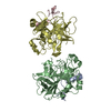 1tq0S S: Starting model for refinement C: citing same article ( |
|---|---|
| Similar structure data |


-Links
 PDBj
PDBj


- Assembly
| Deposited unit | 
| ||||||||
|---|---|---|---|---|---|---|---|---|---|
| 1 | 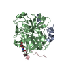
| ||||||||
| 2 | 
| ||||||||
| Unit cell |
| ||||||||
| Details | THE BIOLOGICAL ASSEMBLY CONSISTS OF A, B AND C CHAINS. |
-Components
| #1: Protein/peptide | Mass: 5105.731 Da / Num. of mol.: 2 / Fragment: Light chain: UNP residues 317-360 Source method: isolated from a genetically manipulated source Source: (gene. exp.)  Cricetulus griseus (Chinese hamster) / References: UniProt: P19221, thrombin Cricetulus griseus (Chinese hamster) / References: UniProt: P19221, thrombin#2: Protein | Mass: 29795.461 Da / Num. of mol.: 2 / Fragment: Heavy chain: UNP residues 361-618 / Mutation: W215A, E217A Source method: isolated from a genetically manipulated source Source: (gene. exp.) Cricetulus griseus (Chinese hamster) / References: UniProt: P19221, thrombin#3: Protein/peptide | Mass: 2673.862 Da / Num. of mol.: 2 / Fragment: Extracellular fragment: UNP residues 42-62 Source method: isolated from a genetically manipulated source Source: (gene. exp.) Homo sapiens (human) / Gene: F2R, CF2R, PAR1, TR / Cell (production host): BHK cells / Organ (production host): baby hamster kidney / Production host: Cricetulus griseus (Chinese hamster) / References: UniProt: P25116#4: Sugar |   Type: D-saccharide, beta linking / Mass: 221.208 Da / Num. of mol.: 2 Type: D-saccharide, beta linking / Mass: 221.208 Da / Num. of mol.: 2Source method: isolated from a genetically manipulated source Formula: C8H15NO6 #5: Water | ChemComp-HOH / |  Mass: 18.015 Da / Num. of mol.: 217 / Source method: isolated from a natural source / Formula: H2O Mass: 18.015 Da / Num. of mol.: 217 / Source method: isolated from a natural source / Formula: H2OHas protein modification | Y | |
|---|
-Experimental details
-Experiment
| Experiment | Method: X-RAY DIFFRACTION / Number of used crystals: 1 |
|---|
- Sample preparation
| Crystal | Density Matthews: 3.16 Å3/Da / Density % sol: 61.1 % |
|---|---|
| Crystal grow | Temperature: 295 K / Method: vapor diffusion, hanging drop / pH: 8.5 Details: 100mM Tris-HCl pH 8.5, 20% PEG 10000, VAPOR DIFFUSION, HANGING DROP, temperature 295K |
-Data collection
| Diffraction | Mean temperature: 100 K |
|---|---|
| Diffraction source | Source: SYNCHROTRON / Site: APS  / Beamline: 14-BM-C / Wavelength: 0.9 Å / Beamline: 14-BM-C / Wavelength: 0.9 Å |
| Detector | Type: ADSC QUANTUM 315 / Detector: CCD / Date: Dec 22, 2008 |
| Radiation | Monochromator: Bent Ge(111) / Protocol: SINGLE WAVELENGTH / Monochromatic (M) / Laue (L): M / Scattering type: x-ray |
| Radiation wavelength | Wavelength: 0.9 Å / Relative weight: 1 |
| Reflection | Resolution: 2.2→40 Å / Num. all: 49562 / Num. obs: 45052 / % possible obs: 90.9 % / Observed criterion σ(I): -3 / Redundancy: 6.9 % / Biso Wilson estimate: 20.2 Å2 / Rmerge(I) obs: 0.051 / Net I/σ(I): 28.6 |
| Reflection shell | Resolution: 2.2→2.28 Å / Redundancy: 6.3 % / Rmerge(I) obs: 0.365 / Mean I/σ(I) obs: 4.9 / Num. unique all: 3936 / % possible all: 80.9 |
- Processing
| Software |
| ||||||||||||||||||||||||||||||||||||
|---|---|---|---|---|---|---|---|---|---|---|---|---|---|---|---|---|---|---|---|---|---|---|---|---|---|---|---|---|---|---|---|---|---|---|---|---|---|
| Refinement | Method to determine structure: MOLECULAR REPLACEMENT Starting model: PDB entry 1TQ0 Resolution: 2.2→26.23 Å / Rfactor Rfree error: 0.004 / Data cutoff high absF: 150926.62 / Data cutoff low absF: 0 / Isotropic thermal model: RESTRAINED / Cross valid method: THROUGHOUT / σ(F): 0 / σ(I): 0 / Stereochemistry target values: Engh & Huber
| ||||||||||||||||||||||||||||||||||||
| Displacement parameters | Biso mean: 41.1 Å2 | ||||||||||||||||||||||||||||||||||||
| Refine analyze |
| ||||||||||||||||||||||||||||||||||||
| Refinement step | Cycle: LAST / Resolution: 2.2→26.23 Å
| ||||||||||||||||||||||||||||||||||||
| Refine LS restraints |
| ||||||||||||||||||||||||||||||||||||
| LS refinement shell | Resolution: 2.2→2.34 Å / Rfactor Rfree error: 0.015 / Total num. of bins used: 6
|
