 Movie
Movie Controller

[English] 日本語
 Yorodumi
- PDB-4afv: THE STRUCTURE OF METACASPASE 2 FROM T. BRUCEI DETERMINED IN THE P... -
+ Open data

- Basic information
| Entry | Database: PDB / ID: 4afv | ||||||
|---|---|---|---|---|---|---|---|
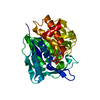
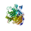
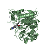
| Title | THE STRUCTURE OF METACASPASE 2 FROM T. BRUCEI DETERMINED IN THE PRESENCE OF CALCIUM CHLORIDE | ||||||
 Components Components | METACASPASE MCA2 | ||||||
 Keywords Keywords | HYDROLASE / CYSTEINE PEPTIDASE / CASPASE/HEMOGLOBIN FOLD | ||||||
| Function / homology |  Function and homology information Function and homology informationcysteine-type peptidase activity / recycling endosome / Hydrolases; Acting on peptide bonds (peptidases); Cysteine endopeptidases / cysteine-type endopeptidase activity / proteolysis / metal ion binding / nucleus / cytoplasm Similarity search - Function | ||||||
| Biological species |  | ||||||
| Method |  X-RAY DIFFRACTION / SYNCHROTRON / MOLECULAR REPLACEMENT / Resolution: 1.5 Å X-RAY DIFFRACTION / SYNCHROTRON / MOLECULAR REPLACEMENT / Resolution: 1.5 Å | ||||||
 Authors Authors | McLuskey, K. / Rudolf, J. / Isaacs, N.W. / Coombs, G.H. / Moss, C.X. / Mottram, J.C. | ||||||
 Citation Citation | Journal: Proc.Natl.Acad.Sci.USA / Year: 2012 Title: Crystal Structure of a Trypanosoma Brucei Metacaspase. Authors: Mcluskey, K. / Rudolf, J. / Proto, W.R. / Isaacs, N.W. / Coombs, G.H. / Moss, C.X. / Mottram, J.C. | ||||||
| History |
|
- Structure visualization
| Structure viewer | Molecule: MolmilJmol/JSmol |
|---|
- Downloads & links
-Download
| PDBx/mmCIF format | 4afv.cif.gz | 153.9 KB | Display | PDBx/mmCIF format |
|---|---|---|---|---|
| PDB format | pdb4afv.ent.gz | 122.6 KB | Display | PDB format |
| PDBx/mmJSON format | 4afv.json.gz | Tree view | PDBx/mmJSON format | |
| Others |  Other downloads Other downloads |
-Validation report
| Arichive directory | https://data.pdbj.org/pub/pdb/validation_reports/af/4afvftp://data.pdbj.org/pub/pdb/validation_reports/af/4afv | HTTPS FTP |
|---|
-Related structure data
| Related structure data | 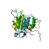 4af8SC  4afpC 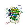 4afrC S: Starting model for refinement C: citing same article ( |
|---|---|
| Similar structure data |


-Links
 PDBj
PDBj

- Assembly
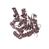
| Deposited unit | 
| |||||||||
|---|---|---|---|---|---|---|---|---|---|---|
| 1 |
| |||||||||
| Unit cell |
| |||||||||
| Components on special symmetry positions |
|
-Components
| #1: Protein | Mass: 39958.348 Da / Num. of mol.: 1 / Mutation: YES Source method: isolated from a genetically manipulated source Source: (gene. exp.)  | ||||
|---|---|---|---|---|---|
| #2: Water | ChemComp-HOH /  Mass: 18.015 Da / Num. of mol.: 287 / Source method: isolated from a natural source / Formula: H2O Mass: 18.015 Da / Num. of mol.: 287 / Source method: isolated from a natural source / Formula: H2O | ||||
| Compound details | ENGINEERED| Has protein modification | Y | Sequence details | UNIPROT Q585F3 DIFFERS FROM THIS ENTRY AT POSITION 315 (GLY-ARG). Q585F3 IS THE NEAREST UNIPROT ...UNIPROT Q585F3 DIFFERS FROM THIS ENTRY AT POSITION 315 (GLY-ARG). Q585F3 IS THE NEAREST UNIPROT ENTRY TO MCA2 FROM T. BRUCEI STRAIN 427. | |
-Experimental details
-Experiment
| Experiment | Method: X-RAY DIFFRACTION / Number of used crystals: 1 |
|---|
- Sample preparation
| Crystal | Density Matthews: 1.87 Å3/Da / Density % sol: 35 % / Description: NONE |
|---|---|
| Crystal grow | pH: 7 Details: 50 MM HEPES PH 7.0, 0.1% TRYPTONE, 20% PEG 3350. CRYSTAL SOAKING FOR 1HR IN 5MM CACL2. |
-Data collection
| Diffraction | Mean temperature: 100 K |
|---|---|
| Diffraction source | Source: SYNCHROTRON / Site: Diamond  / Beamline: I03 / Wavelength: 0.9763 / Beamline: I03 / Wavelength: 0.9763 |
| Detector | Type: ADSC QUANTUM 315r / Detector: CCD / Date: Feb 20, 2011 / Details: MIRRORS |
| Radiation | Monochromator: SI (111) / Protocol: SINGLE WAVELENGTH / Monochromatic (M) / Laue (L): M / Scattering type: x-ray |
| Radiation wavelength | Wavelength: 0.9763 Å / Relative weight: 1 |
| Reflection | Resolution: 1.5→27.38 Å / Num. obs: 45982 / % possible obs: 97.3 % / Observed criterion σ(I): 2 / Redundancy: 3.2 % / Rmerge(I) obs: 0.04 / Net I/σ(I): 16.4 |
| Reflection shell | Resolution: 1.5→1.58 Å / Redundancy: 2.7 % / Rmerge(I) obs: 0.13 / Mean I/σ(I) obs: 6.1 / % possible all: 91 |
- Processing
| Software |
| ||||||||||||||||||||||||||||||||||||||||||||||||||||||||||||||||||||||||||||||||||||||||||||||||||||||||||||||||||||||||||||||||||||||||||||||||||||||||||||||||||||||||||||||||||||||
|---|---|---|---|---|---|---|---|---|---|---|---|---|---|---|---|---|---|---|---|---|---|---|---|---|---|---|---|---|---|---|---|---|---|---|---|---|---|---|---|---|---|---|---|---|---|---|---|---|---|---|---|---|---|---|---|---|---|---|---|---|---|---|---|---|---|---|---|---|---|---|---|---|---|---|---|---|---|---|---|---|---|---|---|---|---|---|---|---|---|---|---|---|---|---|---|---|---|---|---|---|---|---|---|---|---|---|---|---|---|---|---|---|---|---|---|---|---|---|---|---|---|---|---|---|---|---|---|---|---|---|---|---|---|---|---|---|---|---|---|---|---|---|---|---|---|---|---|---|---|---|---|---|---|---|---|---|---|---|---|---|---|---|---|---|---|---|---|---|---|---|---|---|---|---|---|---|---|---|---|---|---|---|---|
| Refinement | Method to determine structure: MOLECULAR REPLACEMENT Starting model: PDB ENTRY 4AF8 Resolution: 1.5→76.1 Å / Cor.coef. Fo:Fc: 0.967 / Cor.coef. Fo:Fc free: 0.953 / SU B: 2.246 / SU ML: 0.039 / Cross valid method: THROUGHOUT / ESU R: 0.098 / ESU R Free: 0.074 / Stereochemistry target values: MAXIMUM LIKELIHOOD Details: HYDROGENS HAVE BEEN ADDED IN THE RIDING POSITIONS. RESIDUES 1-14, 166-171 AND 267-276 ARE DISORDERED.
| ||||||||||||||||||||||||||||||||||||||||||||||||||||||||||||||||||||||||||||||||||||||||||||||||||||||||||||||||||||||||||||||||||||||||||||||||||||||||||||||||||||||||||||||||||||||
| Solvent computation | Ion probe radii: 0.8 Å / Shrinkage radii: 0.08 Å / VDW probe radii: 1.4 Å / Solvent model: BULK SOLVENT MODELLING WITH MASK | ||||||||||||||||||||||||||||||||||||||||||||||||||||||||||||||||||||||||||||||||||||||||||||||||||||||||||||||||||||||||||||||||||||||||||||||||||||||||||||||||||||||||||||||||||||||
| Displacement parameters | Biso mean: 16.55 Å2
| ||||||||||||||||||||||||||||||||||||||||||||||||||||||||||||||||||||||||||||||||||||||||||||||||||||||||||||||||||||||||||||||||||||||||||||||||||||||||||||||||||||||||||||||||||||||
| Refinement step | Cycle: LAST / Resolution: 1.5→76.1 Å
| ||||||||||||||||||||||||||||||||||||||||||||||||||||||||||||||||||||||||||||||||||||||||||||||||||||||||||||||||||||||||||||||||||||||||||||||||||||||||||||||||||||||||||||||||||||||
| Refine LS restraints |
|