 Movie
Movie Controller

[English] 日本語
 Yorodumi
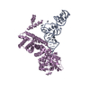
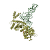
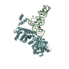
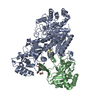
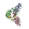
- PDB-3hay: Crystal structure of a substrate-bound full H/ACA RNP from Pyroco... -
+ Open data

- Basic information
| Entry | Database: PDB / ID: 3hay | ||||||
|---|---|---|---|---|---|---|---|
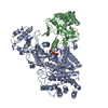
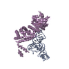
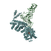
| Title | Crystal structure of a substrate-bound full H/ACA RNP from Pyrococcus furiosus | ||||||
 Components Components |
| ||||||
 Keywords Keywords | ISOMERASE/BIOSYNTHETIC PROTEIN/RNA / H/ACA / guide RNA / RNA-protein complex / pseudouridine synthase / Isomerase / tRNA processing / Ribonucleoprotein / Ribosome biogenesis / rRNA processing / Ribosomal protein / RNA-binding / ISOMERASE-BIOSYNTHETIC PROTEIN-RNA COMPLEX | ||||||
| Function / homology |  Function and homology information Function and homology informationtRNA pseudouridine55 synthase / tRNA pseudouridine(55) synthase activity / pseudouridine synthesis / rRNA pseudouridine synthesis / box H/ACA sno(s)RNA 3'-end processing / tRNA pseudouridine synthesis / snRNA pseudouridine synthesis / mRNA pseudouridine synthesis / ribonuclease P activity / tRNA 5'-leader removal ...tRNA pseudouridine55 synthase / tRNA pseudouridine(55) synthase activity / pseudouridine synthesis / rRNA pseudouridine synthesis / box H/ACA sno(s)RNA 3'-end processing / tRNA pseudouridine synthesis / snRNA pseudouridine synthesis / mRNA pseudouridine synthesis / ribonuclease P activity / tRNA 5'-leader removal / snoRNA binding / rRNA processing / ribosome biogenesis / rRNA binding / structural constituent of ribosome / ribosome / translation / ribonucleoprotein complex / RNA binding / cytoplasm Similarity search - Function | ||||||
| Biological species |   Pyrococcus furiosus (archaea) Pyrococcus furiosus (archaea) | ||||||
| Method |  X-RAY DIFFRACTION / SYNCHROTRON / MOLECULAR REPLACEMENT / Resolution: 4.99 Å X-RAY DIFFRACTION / SYNCHROTRON / MOLECULAR REPLACEMENT / Resolution: 4.99 Å | ||||||
 Authors Authors | Ye, K. | ||||||
 Citation Citation | Journal: Mol.Cell / Year: 2009 Title: Structural mechanism of substrate RNA recruitment in H/ACA RNA-guided pseudouridine synthase. Authors: Duan, J. / Li, L. / Lu, J. / Wang, W. / Ye, K. | ||||||
| History |
|
- Structure visualization
| Structure viewer | Molecule: MolmilJmol/JSmol |
|---|
- Downloads & links
-Download
| PDBx/mmCIF format | 3hay.cif.gz | 171.5 KB | Display | PDBx/mmCIF format |
|---|---|---|---|---|
| PDB format | pdb3hay.ent.gz | 129.3 KB | Display | PDB format |
| PDBx/mmJSON format | 3hay.json.gz | Tree view | PDBx/mmJSON format | |
| Others |  Other downloads Other downloads |
-Validation report
| Arichive directory | https://data.pdbj.org/pub/pdb/validation_reports/ha/3hayftp://data.pdbj.org/pub/pdb/validation_reports/ha/3hay | HTTPS FTP |
|---|
-Related structure data
| Related structure data | 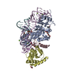 3haxSC S: Starting model for refinement C: citing same article ( |
|---|---|
| Similar structure data |


-Links
 PDBj
PDBj


- Assembly
| Deposited unit | 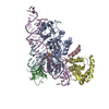
| ||||||||
|---|---|---|---|---|---|---|---|---|---|
| 1 |
| ||||||||
| Unit cell |
|
-Components
-Protein , 4 types, 4 molecules ABCD
| #1: Protein | Mass: 39453.898 Da / Num. of mol.: 1 Source method: isolated from a genetically manipulated source Source: (gene. exp.) Pyrococcus furiosus (archaea) / Gene: truB, PF1785 / Production host:  References: UniProt: Q7LWY0, Isomerases; Intramolecular transferases; Transferring other groups |
|---|---|
| #2: Protein | Mass: 12340.823 Da / Num. of mol.: 1 Source method: isolated from a genetically manipulated source Source: (gene. exp.) Pyrococcus furiosus (archaea) / Gene: PF1791 / Production host: |
| #3: Protein | Mass: 7214.603 Da / Num. of mol.: 1 Source method: isolated from a genetically manipulated source Source: (gene. exp.) Pyrococcus furiosus (archaea) / Gene: PF1141 / Production host: |
| #4: Protein | Mass: 14314.623 Da / Num. of mol.: 1 Source method: isolated from a genetically manipulated source Source: (gene. exp.) Pyrococcus furiosus (archaea) / Gene: rpl7ae, PF1367 / Production host: |
-RNA chain , 2 types, 2 molecules EF
| #5: RNA chain | Mass: 22949.637 Da / Num. of mol.: 1 / Source method: obtained synthetically / Details: RNA WAS PREPARED BY IN VITRO TRANSCRIPTION |
|---|---|
| #6: RNA chain | Mass: 4452.682 Da / Num. of mol.: 1 / Source method: obtained synthetically |
-Non-polymers , 1 types, 1 molecules 
| #7: Chemical | ChemComp-ZN /  Mass: 65.409 Da / Num. of mol.: 1 / Source method: obtained synthetically / Formula: Zn Mass: 65.409 Da / Num. of mol.: 1 / Source method: obtained synthetically / Formula: Zn |
|---|
-Experimental details
-Experiment
| Experiment | Method: X-RAY DIFFRACTION / Number of used crystals: 1 |
|---|
- Sample preparation
| Crystal grow | Temperature: 293 K / Method: vapor diffusion, hanging drop / pH: 4.9 Details: 1.5M lithium sulfate, 50mM sodium acetate (pH 4.9), VAPOR DIFFUSION, HANGING DROP, temperature 293K |
|---|
-Data collection
| Diffraction | Mean temperature: 100 K |
|---|---|
| Diffraction source | Source: SYNCHROTRON / Site: SPring-8  / Beamline: BL41XU / Wavelength: 1 Å / Beamline: BL41XU / Wavelength: 1 Å |
| Detector | Date: Nov 15, 2006 |
| Radiation | Protocol: SINGLE WAVELENGTH / Monochromatic (M) / Laue (L): M / Scattering type: x-ray |
| Radiation wavelength | Wavelength: 1 Å / Relative weight: 1 |
| Reflection | Resolution: 4.99→50 Å / Num. obs: 13408 / % possible obs: 99.5 % / Redundancy: 10.2 % / Rmerge(I) obs: 0.082 / Net I/σ(I): 34.4 |
| Reflection shell | Resolution: 5→5.18 Å / Rmerge(I) obs: 0.481 / Mean I/σ(I) obs: 4.7 / % possible all: 99.8 |
- Processing
| Software |
| ||||||||||||||||||||||||||||||||||||||||||||||||||||||||||||||||||||||||||||||||||||||||||||||||||||||||||||||||||||||||||||||||||||||||||||||||||||||||||||||||||||||||||
|---|---|---|---|---|---|---|---|---|---|---|---|---|---|---|---|---|---|---|---|---|---|---|---|---|---|---|---|---|---|---|---|---|---|---|---|---|---|---|---|---|---|---|---|---|---|---|---|---|---|---|---|---|---|---|---|---|---|---|---|---|---|---|---|---|---|---|---|---|---|---|---|---|---|---|---|---|---|---|---|---|---|---|---|---|---|---|---|---|---|---|---|---|---|---|---|---|---|---|---|---|---|---|---|---|---|---|---|---|---|---|---|---|---|---|---|---|---|---|---|---|---|---|---|---|---|---|---|---|---|---|---|---|---|---|---|---|---|---|---|---|---|---|---|---|---|---|---|---|---|---|---|---|---|---|---|---|---|---|---|---|---|---|---|---|---|---|---|---|---|---|---|
| Refinement | Method to determine structure: MOLECULAR REPLACEMENT Starting model: 3HAX Resolution: 4.99→20 Å / Cor.coef. Fo:Fc: 0.891 / Cor.coef. Fo:Fc free: 0.873 / SU B: 127.599 / SU ML: 1.398 / Cross valid method: THROUGHOUT / σ(F): 0 / ESU R Free: 1.323 / Stereochemistry target values: Engh & Huber / Details: HYDROGENS HAVE BEEN ADDED IN THE RIDING POSITIONS
| ||||||||||||||||||||||||||||||||||||||||||||||||||||||||||||||||||||||||||||||||||||||||||||||||||||||||||||||||||||||||||||||||||||||||||||||||||||||||||||||||||||||||||
| Solvent computation | Ion probe radii: 0.8 Å / Shrinkage radii: 0.8 Å / VDW probe radii: 1.4 Å / Solvent model: MASK | ||||||||||||||||||||||||||||||||||||||||||||||||||||||||||||||||||||||||||||||||||||||||||||||||||||||||||||||||||||||||||||||||||||||||||||||||||||||||||||||||||||||||||
| Displacement parameters | Biso mean: 263.788 Å2
| ||||||||||||||||||||||||||||||||||||||||||||||||||||||||||||||||||||||||||||||||||||||||||||||||||||||||||||||||||||||||||||||||||||||||||||||||||||||||||||||||||||||||||
| Refinement step | Cycle: LAST / Resolution: 4.99→20 Å
| ||||||||||||||||||||||||||||||||||||||||||||||||||||||||||||||||||||||||||||||||||||||||||||||||||||||||||||||||||||||||||||||||||||||||||||||||||||||||||||||||||||||||||
| Refine LS restraints |
| ||||||||||||||||||||||||||||||||||||||||||||||||||||||||||||||||||||||||||||||||||||||||||||||||||||||||||||||||||||||||||||||||||||||||||||||||||||||||||||||||||||||||||
| LS refinement shell | Resolution: 4.992→5.113 Å / Total num. of bins used: 20
|
