 Movie
Movie Controller


+ Open data

- Basic information
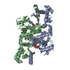
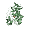
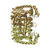
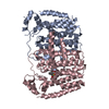
| Entry | Database: PDB / ID: 3h9g | ||||||
|---|---|---|---|---|---|---|---|
| Title | Crystal structure of E. coli MccB + MccA-N7isoASN | ||||||
 Components Components |
| ||||||
 Keywords Keywords | TRANSFERASE/ANTIBIOTIC / Ubiquitin-activating enzyme / microcin / bacteriocin / Mcc7 / peptide antibiotic / N-P bond formation / Antibiotic / Antimicrobial / Phosphoprotein / TRANSFERASE / TRANSFERASE-ANTIBIOTIC COMPLEX | ||||||
| Function / homology |  Function and homology information Function and homology informationubiquitin-like modifier activating enzyme activity / thiosulfate-cyanide sulfurtransferase activity / nucleotidyltransferase activity / defense response to bacterium / ATP binding / metal ion binding / cytoplasm Similarity search - Function | ||||||
| Biological species |  | ||||||
| Method |  X-RAY DIFFRACTION / SYNCHROTRON / MOLECULAR REPLACEMENT / Resolution: 2.2 Å X-RAY DIFFRACTION / SYNCHROTRON / MOLECULAR REPLACEMENT / Resolution: 2.2 Å | ||||||
 Authors Authors | Regni, C.A. / Roush, R.F. / Miller, D. / Nourse, A. / Walsh, C.T. / Schulman, B.A. | ||||||
 Citation Citation | Journal: Embo J. / Year: 2009 Title: How the MccB bacterial ancestor of ubiquitin E1 initiates biosynthesis of the microcin C7 antibiotic. Authors: Regni, C.A. / Roush, R.F. / Miller, D.J. / Nourse, A. / Walsh, C.T. / Schulman, B.A. | ||||||
| History |
|
- Structure visualization
| Structure viewer | Molecule: MolmilJmol/JSmol |
|---|
- Downloads & links
-Download
| PDBx/mmCIF format | 3h9g.cif.gz | 285.6 KB | Display | PDBx/mmCIF format |
|---|---|---|---|---|
| PDB format | pdb3h9g.ent.gz | 229.4 KB | Display | PDB format |
| PDBx/mmJSON format | 3h9g.json.gz | Tree view | PDBx/mmJSON format | |
| Others |  Other downloads Other downloads |
-Validation report
| Arichive directory | https://data.pdbj.org/pub/pdb/validation_reports/h9/3h9gftp://data.pdbj.org/pub/pdb/validation_reports/h9/3h9g | HTTPS FTP |
|---|
-Related structure data
| Related structure data | 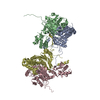 3h5aSC 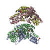 3h5nC 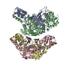 3h5rC  3h9jC 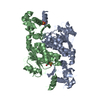 3h9qC S: Starting model for refinement C: citing same article ( |
|---|---|
| Similar structure data |


-Links
 PDBj
PDBj

- Assembly
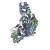
| Deposited unit | 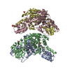
| ||||||||
|---|---|---|---|---|---|---|---|---|---|
| 1 | 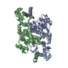
| ||||||||
| 2 | 
| ||||||||
| Unit cell |
|
-Components
| #1: Protein | Mass: 39409.766 Da / Num. of mol.: 4 Source method: isolated from a genetically manipulated source Details: thrombin cleavable His-tag / Source: (gene. exp.) #2: Protein/peptide | 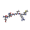  Type: Polypeptide / Class: Inhibitor / Mass: 763.844 Da / Num. of mol.: 4 / Mutation: ASN 7 to XSN, Iso asparagine / Source method: obtained synthetically Type: Polypeptide / Class: Inhibitor / Mass: 763.844 Da / Num. of mol.: 4 / Mutation: ASN 7 to XSN, Iso asparagine / Source method: obtained syntheticallyDetails: MRTGNA-N7isoAsn was synthesized as previously described (Novoa et al, 1986; Roush et. al. 2008) Source: (synth.) #3: Chemical | ChemComp-ZN /   Mass: 65.409 Da / Num. of mol.: 4 / Source method: obtained synthetically / Formula: Zn Mass: 65.409 Da / Num. of mol.: 4 / Source method: obtained synthetically / Formula: Zn#4: Chemical | ChemComp-SO4 /   Mass: 96.063 Da / Num. of mol.: 7 / Source method: obtained synthetically / Formula: SO4 Mass: 96.063 Da / Num. of mol.: 7 / Source method: obtained synthetically / Formula: SO4#5: Water | ChemComp-HOH / |  Mass: 18.015 Da / Num. of mol.: 511 / Source method: isolated from a natural source / Formula: H2O Mass: 18.015 Da / Num. of mol.: 511 / Source method: isolated from a natural source / Formula: H2O |
|---|
-Experimental details
-Experiment
| Experiment | Method: X-RAY DIFFRACTION / Number of used crystals: 1 |
|---|
- Sample preparation
| Crystal | Density Matthews: 1.94 Å3/Da / Density % sol: 36.66 % |
|---|---|
| Crystal grow | Temperature: 291.2 K / Method: vapor diffusion, hanging drop / pH: 8 Details: 24-26% Pentaerythritol ethoxylate (15/4 EO/OH, Hampton Research), 50 mM Tris-HCl pH 8.0, 50 mM (NH4)2SO4, 10 mM MccA-N7isoAsn, VAPOR DIFFUSION, HANGING DROP, temperature 291.2K |
-Data collection
| Diffraction | Mean temperature: 100 K |
|---|---|
| Diffraction source | Source: SYNCHROTRON / Site: APS  / Beamline: 22-ID / Wavelength: 0.97926 Å / Beamline: 22-ID / Wavelength: 0.97926 Å |
| Detector | Type: MAR scanner 300 mm plate / Detector: IMAGE PLATE / Date: Dec 2, 2007 / Details: Rosenbaum-Rock vertical focusing mirror |
| Radiation | Monochromator: Rosenbaum-Rock high-resolution double crystal Si(220) sagital focusing Protocol: SINGLE WAVELENGTH / Monochromatic (M) / Laue (L): M / Scattering type: x-ray |
| Radiation wavelength | Wavelength: 0.97926 Å / Relative weight: 1 |
| Reflection | Resolution: 2.2→50 Å / Num. all: 60798 / Num. obs: 60798 / % possible obs: 96.7 % / Observed criterion σ(F): 0 / Observed criterion σ(I): 0 / Redundancy: 4.1 % / Biso Wilson estimate: 33.6 Å2 / Rmerge(I) obs: 0.065 / Χ2: 1.152 / Net I/σ(I): 21.914 |
| Reflection shell | Resolution: 2.2→2.28 Å / Redundancy: 3.6 % / Rmerge(I) obs: 0.371 / Num. unique all: 5645 / Χ2: 0.822 / % possible all: 90.5 |
- Processing
| Software |
| ||||||||||||||||||||||||||||||||||||||||||||||||||||||||||||||||||||||||||||||||||||||||||
|---|---|---|---|---|---|---|---|---|---|---|---|---|---|---|---|---|---|---|---|---|---|---|---|---|---|---|---|---|---|---|---|---|---|---|---|---|---|---|---|---|---|---|---|---|---|---|---|---|---|---|---|---|---|---|---|---|---|---|---|---|---|---|---|---|---|---|---|---|---|---|---|---|---|---|---|---|---|---|---|---|---|---|---|---|---|---|---|---|---|---|---|
| Refinement | Method to determine structure: MOLECULAR REPLACEMENT Starting model: PDB entry 3H5A Resolution: 2.2→46.88 Å / Cor.coef. Fo:Fc: 0.946 / Cor.coef. Fo:Fc free: 0.908 / WRfactor Rfree: 0.249 / WRfactor Rwork: 0.194 / Occupancy max: 1 / Occupancy min: 1 / FOM work R set: 0.799 / SU B: 7.969 / SU ML: 0.202 / SU R Cruickshank DPI: 0.451 / SU Rfree: 0.263 / Cross valid method: THROUGHOUT / σ(I): 0 / ESU R: 0.451 / ESU R Free: 0.263 / Stereochemistry target values: MAXIMUM LIKELIHOOD / Details: HYDROGENS HAVE BEEN ADDED IN THE RIDING POSITIONS
| ||||||||||||||||||||||||||||||||||||||||||||||||||||||||||||||||||||||||||||||||||||||||||
| Solvent computation | Ion probe radii: 0.8 Å / Shrinkage radii: 0.8 Å / VDW probe radii: 1.2 Å / Solvent model: MASK | ||||||||||||||||||||||||||||||||||||||||||||||||||||||||||||||||||||||||||||||||||||||||||
| Displacement parameters | Biso max: 78.59 Å2 / Biso mean: 36.297 Å2 / Biso min: 15.17 Å2
| ||||||||||||||||||||||||||||||||||||||||||||||||||||||||||||||||||||||||||||||||||||||||||
| Refinement step | Cycle: LAST / Resolution: 2.2→46.88 Å
| ||||||||||||||||||||||||||||||||||||||||||||||||||||||||||||||||||||||||||||||||||||||||||
| Refine LS restraints |
| ||||||||||||||||||||||||||||||||||||||||||||||||||||||||||||||||||||||||||||||||||||||||||
| LS refinement shell | Resolution: 2.2→2.257 Å / Total num. of bins used: 20
|
