 Movie
Movie Controller


+ Open data

- Basic information
| Entry | Database: PDB / ID: 3h6x | ||||||
|---|---|---|---|---|---|---|---|
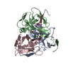
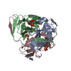
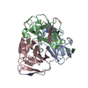
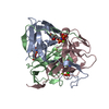
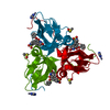
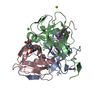
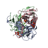
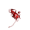
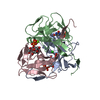
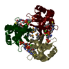
| Title | Crystal structure of dUTPase from Streptococcus mutans | ||||||
 Components Components | dUTPase | ||||||
 Keywords Keywords | HYDROLASE / jelly-roll beta-barrel | ||||||
| Function / homology |  Function and homology information Function and homology informationdUTP catabolic process / dUMP biosynthetic process / dUTP diphosphatase / dUTP diphosphatase activity / magnesium ion binding Similarity search - Function | ||||||
| Biological species |  Streptococcus mutans (bacteria) Streptococcus mutans (bacteria) | ||||||
| Method |  X-RAY DIFFRACTION / SYNCHROTRON / SAD / Resolution: 1.7 Å X-RAY DIFFRACTION / SYNCHROTRON / SAD / Resolution: 1.7 Å | ||||||
 Authors Authors | Li, G.L. / Wang, K.T. / Liu, X. / Li, L.F. / Su, X.D. | ||||||
 Citation Citation | Journal: To be Published Title: Structure and activity analysis of dUTP nucleotidohydrolase from Streptococcus mutans Authors: Li, G.L. / Wang, K.T. / Liu, X. / Li, L.F. / Su, X.D. | ||||||
| History |
|
- Structure visualization
| Structure viewer | Molecule: MolmilJmol/JSmol |
|---|
- Downloads & links
-Download
| PDBx/mmCIF format | 3h6x.cif.gz | 102.5 KB | Display | PDBx/mmCIF format |
|---|---|---|---|---|
| PDB format | pdb3h6x.ent.gz | 78.9 KB | Display | PDB format |
| PDBx/mmJSON format | 3h6x.json.gz | Tree view | PDBx/mmJSON format | |
| Others |  Other downloads Other downloads |
-Validation report
| Arichive directory | https://data.pdbj.org/pub/pdb/validation_reports/h6/3h6xftp://data.pdbj.org/pub/pdb/validation_reports/h6/3h6x | HTTPS FTP |
|---|
-Related structure data
| Similar structure data |
|---|
-Links
 PDBj
PDBj
- Assembly
| Deposited unit | 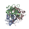
| ||||||||||||||||||||||||||||||||||||
|---|---|---|---|---|---|---|---|---|---|---|---|---|---|---|---|---|---|---|---|---|---|---|---|---|---|---|---|---|---|---|---|---|---|---|---|---|---|
| 1 |
| ||||||||||||||||||||||||||||||||||||
| Unit cell |
| ||||||||||||||||||||||||||||||||||||
| Components on special symmetry positions |
| ||||||||||||||||||||||||||||||||||||
| Noncrystallographic symmetry (NCS) | NCS domain:
NCS domain segments:
|
-Components
| #1: Protein | Mass: 16391.641 Da / Num. of mol.: 3 Source method: isolated from a genetically manipulated source Source: (gene. exp.) Streptococcus mutans (bacteria) / Gene: dut / Plasmid: pET28a / Production host: #2: Water | ChemComp-HOH / |  Mass: 18.015 Da / Num. of mol.: 688 / Source method: isolated from a natural source / Formula: H2O Mass: 18.015 Da / Num. of mol.: 688 / Source method: isolated from a natural source / Formula: H2OHas protein modification | Y | |
|---|
-Experimental details
-Experiment
| Experiment | Method: X-RAY DIFFRACTION / Number of used crystals: 1 |
|---|
- Sample preparation
| Crystal | Density Matthews: 2.07 Å3/Da / Density % sol: 40.54 % |
|---|---|
| Crystal grow | Temperature: 289 K / Method: vapor diffusion, sitting drop / pH: 7.5 Details: 0.2M MgCl2, 25% polyethylene glycol(PEG)3350, 0.1M Tris-HCl(pH 8.5), pH7.5, VAPOR DIFFUSION, SITTING DROP, temperature 289K |
-Data collection
| Diffraction | Mean temperature: 100 K |
|---|---|
| Diffraction source | Source: SYNCHROTRON / Site: MAX II  / Beamline: I911-3 / Wavelength: 0.9787 Å / Beamline: I911-3 / Wavelength: 0.9787 Å |
| Detector | Type: MAR CCD 165 mm / Detector: CCD / Date: May 25, 2008 |
| Radiation | Protocol: SINGLE WAVELENGTH / Monochromatic (M) / Laue (L): M / Scattering type: x-ray |
| Radiation wavelength | Wavelength: 0.9787 Å / Relative weight: 1 |
| Reflection | Resolution: 1.7→30 Å / Num. obs: 43830 / % possible obs: 98.6 % / Observed criterion σ(I): -3 / Redundancy: 3.6 % / Biso Wilson estimate: 11.33 Å2 / Rsym value: 0.047 / Net I/σ(I): 15.25 |
| Reflection shell | Resolution: 1.7→1.8 Å / Redundancy: 3.5 % / Mean I/σ(I) obs: 8.97 / Num. unique all: 7142 / Rsym value: 0.123 / % possible all: 96.1 |
- Processing
| Software |
| |||||||||||||||||||||||||||||||||||||||||||||||||||||||||||||||||||||||||||||||||||||||||||||||||||||||||
|---|---|---|---|---|---|---|---|---|---|---|---|---|---|---|---|---|---|---|---|---|---|---|---|---|---|---|---|---|---|---|---|---|---|---|---|---|---|---|---|---|---|---|---|---|---|---|---|---|---|---|---|---|---|---|---|---|---|---|---|---|---|---|---|---|---|---|---|---|---|---|---|---|---|---|---|---|---|---|---|---|---|---|---|---|---|---|---|---|---|---|---|---|---|---|---|---|---|---|---|---|---|---|---|---|---|---|
| Refinement | Method to determine structure: SAD / Resolution: 1.7→19.941 Å / Cor.coef. Fo:Fc: 0.952 / Cor.coef. Fo:Fc free: 0.93 / Occupancy max: 1 / Occupancy min: 0.7 / FOM work R set: 0.885 / SU B: 1.831 / SU ML: 0.2 / Cross valid method: THROUGHOUT / σ(F): 1.09 / Phase error: 18.8 / Stereochemistry target values: MLHL / Details: HYDROGENS HAVE BEEN ADDED IN THE RIDING POSITIONS
| |||||||||||||||||||||||||||||||||||||||||||||||||||||||||||||||||||||||||||||||||||||||||||||||||||||||||
| Solvent computation | Shrinkage radii: 0.9 Å / VDW probe radii: 1.11 Å / Solvent model: FLAT BULK SOLVENT MODEL / Bsol: 49.132 Å2 / ksol: 0.372 e/Å3 | |||||||||||||||||||||||||||||||||||||||||||||||||||||||||||||||||||||||||||||||||||||||||||||||||||||||||
| Displacement parameters | Biso max: 50.15 Å2 / Biso mean: 14.753 Å2 / Biso min: 1.54 Å2
| |||||||||||||||||||||||||||||||||||||||||||||||||||||||||||||||||||||||||||||||||||||||||||||||||||||||||
| Refinement step | Cycle: LAST / Resolution: 1.7→19.941 Å
| |||||||||||||||||||||||||||||||||||||||||||||||||||||||||||||||||||||||||||||||||||||||||||||||||||||||||
| Refine LS restraints |
| |||||||||||||||||||||||||||||||||||||||||||||||||||||||||||||||||||||||||||||||||||||||||||||||||||||||||
| Refine LS restraints NCS |
| |||||||||||||||||||||||||||||||||||||||||||||||||||||||||||||||||||||||||||||||||||||||||||||||||||||||||
| LS refinement shell |
|
