cathepsin V / RUNX1 regulates transcription of genes involved in differentiation of keratinocytes / Trafficking and processing of endosomal TLR / Assembly of collagen fibrils and other multimeric structures / Activation of Matrix Metalloproteinases / extracellular matrix disassembly / cysteine-type endopeptidase inhibitor activity / Degradation of the extracellular matrix / cysteine-type peptidase activity / MHC class II antigen presentation ...cathepsin V / RUNX1 regulates transcription of genes involved in differentiation of keratinocytes / Trafficking and processing of endosomal TLR / Assembly of collagen fibrils and other multimeric structures / Activation of Matrix Metalloproteinases / extracellular matrix disassembly / cysteine-type endopeptidase inhibitor activity / Degradation of the extracellular matrix / cysteine-type peptidase activity / MHC class II antigen presentation / lysosomal lumen / : / Endosomal/Vacuolar pathway / antigen processing and presentation of exogenous peptide antigen via MHC class II / lysosome / serine-type endopeptidase activity / cysteine-type endopeptidase activity / : / extracellular region Similarity search - Function
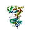
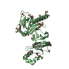
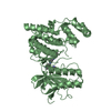
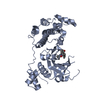
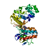
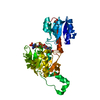
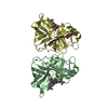
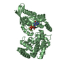
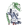
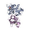
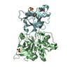
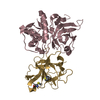
Proteinase inhibitor I48, clitocypin / Peptidase inhibitor clitocypin / Cathepsin propeptide inhibitor domain (I29) / Cathepsin propeptide inhibitor domain (I29) / Cathepsin propeptide inhibitor domain (I29) / Papain-like cysteine endopeptidase / Cysteine peptidase, asparagine active site / Eukaryotic thiol (cysteine) proteases asparagine active site. / Cysteine peptidase, histidine active site / Eukaryotic thiol (cysteine) proteases histidine active site. ...Proteinase inhibitor I48, clitocypin / Peptidase inhibitor clitocypin / Cathepsin propeptide inhibitor domain (I29) / Cathepsin propeptide inhibitor domain (I29) / Cathepsin propeptide inhibitor domain (I29) / Papain-like cysteine endopeptidase / Cysteine peptidase, asparagine active site / Eukaryotic thiol (cysteine) proteases asparagine active site. / Cysteine peptidase, histidine active site / Eukaryotic thiol (cysteine) proteases histidine active site. / : / Peptidase C1A, papain C-terminal / Papain family cysteine protease / Papain family cysteine protease / Cysteine proteinases / Cysteine peptidase, cysteine active site / Eukaryotic thiol (cysteine) proteases cysteine active site. / Cathepsin B; Chain A / Trefoil (Acidic Fibroblast Growth Factor, subunit A) - #50 / Trefoil (Acidic Fibroblast Growth Factor, subunit A) / Trefoil / Papain-like cysteine peptidase superfamily / Alpha-Beta Complex / Mainly Beta / Alpha Beta Similarity search - Domain/homology
Mass: 18.015 Da / Num. of mol.: 857 / Source method: isolated from a natural source / Formula: H2O
Compound details
RESIDUE 25(CYC) IN CATHEPSIN L2 IS MODIFIED. CATHEPSIN L2 WAS TREATED WITH METHYL ...RESIDUE 25(CYC) IN CATHEPSIN L2 IS MODIFIED. CATHEPSIN L2 WAS TREATED WITH METHYL METHANETHIOSULFONATE (MMTS) TO FORM SCH (S-METHYL-THIO-CYSTEINE).
-
Experimental details
-
Experiment
Experiment
Method: X-RAY DIFFRACTION / Number of used crystals: 1
-
Sample preparation
Crystal
Density Matthews: 2.54 Å3/Da / Density % sol: 51.49 %
Crystal grow
Temperature: 293 K / Method: vapor diffusion, sitting drop / pH: 0 Details: 0.4 M Li2SO4, 12% PEG800, 20% glycerol, VAPOR DIFFUSION, SITTING DROP, temperature 293K
In the structure databanks used in Yorodumi, some data are registered as the other names, "COVID-19 virus" and "2019-nCoV". Here are the details of the virus and the list of structure data.
Jan 31, 2019. EMDB accession codes are about to change! (news from PDBe EMDB page)
EMDB accession codes are about to change! (news from PDBe EMDB page)
The allocation of 4 digits for EMDB accession codes will soon come to an end. Whilst these codes will remain in use, new EMDB accession codes will include an additional digit and will expand incrementally as the available range of codes is exhausted. The current 4-digit format prefixed with “EMD-” (i.e. EMD-XXXX) will advance to a 5-digit format (i.e. EMD-XXXXX), and so on. It is currently estimated that the 4-digit codes will be depleted around Spring 2019, at which point the 5-digit format will come into force.
The EM Navigator/Yorodumi systems omit the EMD- prefix.
Related info.:Q: What is EMD? / ID/Accession-code notation in Yorodumi/EM Navigator
Yorodumi is a browser for structure data from EMDB, PDB, SASBDB, etc.
This page is also the successor to EM Navigator detail page, and also detail information page/front-end page for Omokage search.
The word "yorodu" (or yorozu) is an old Japanese word meaning "ten thousand". "mi" (miru) is to see.
Related info.:EMDB / PDB / SASBDB / Comparison of 3 databanks / Yorodumi Search / Aug 31, 2016. New EM Navigator & Yorodumi / Yorodumi Papers / Jmol/JSmol / Function and homology information / Changes in new EM Navigator and Yorodumi
 Movie
Movie Controller


 Open data
 Basic information
 Components
 Keywords
 Function and homology information
 Homo sapiens (human)
 Clitocybe nebularis (fungus)
 X-RAY DIFFRACTION /
  Authors
 Citation
 Structure visualization
 Downloads & links
 Other downloads


 PDBj
PDBj


 Assembly

 Mass: 96.063 Da / Num. of mol.: 6 / Source method: obtained synthetically / Formula: SO4
 Mass: 18.015 Da / Num. of mol.: 857 / Source method: isolated from a natural source / Formula: H2O
 Sample preparation
 / Beamline: 5.2R / Wavelength: 1.1 Å
 Processing