 Movie
Movie Controller

[English] 日本語
 Yorodumi
- PDB-5d1k: Crystal Structure of UbcH5B in Complex with the RING-U5BR Fragmen... -
+ Open data

- Basic information
| Entry | Database: PDB / ID: 5d1k | ||||||
|---|---|---|---|---|---|---|---|
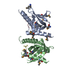
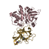
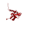
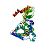
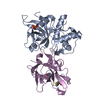
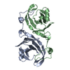
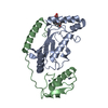
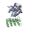
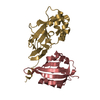
| Title | Crystal Structure of UbcH5B in Complex with the RING-U5BR Fragment of AO7 | ||||||
 Components Components |
| ||||||
 Keywords Keywords | LIGASE / ubiquitin conjugating enzyme (E2) / ubiquitin ligase (E3) / RING finger / ubiquitination / ubiquitin | ||||||
| Function / homology |  Function and homology information Function and homology informationprotein-RNA covalent cross-linking repair / (E3-independent) E2 ubiquitin-conjugating enzyme / protein K6-linked ubiquitination / E2 ubiquitin-conjugating enzyme / ubiquitin conjugating enzyme activity / NF-kappaB binding / protein autoubiquitination / protein K48-linked ubiquitination / rescue of stalled cytosolic ribosome / cytosolic ribosome ...protein-RNA covalent cross-linking repair / (E3-independent) E2 ubiquitin-conjugating enzyme / protein K6-linked ubiquitination / E2 ubiquitin-conjugating enzyme / ubiquitin conjugating enzyme activity / NF-kappaB binding / protein autoubiquitination / protein K48-linked ubiquitination / rescue of stalled cytosolic ribosome / cytosolic ribosome / : / protein modification process / Synthesis of active ubiquitin: roles of E1 and E2 enzymes / TICAM1, RIP1-mediated IKK complex recruitment / IKK complex recruitment mediated by RIP1 / PINK1-PRKN Mediated Mitophagy / Negative regulators of DDX58/IFIH1 signaling / Peroxisomal protein import / Regulation of TNFR1 signaling / RING-type E3 ubiquitin transferase / Inactivation of CSF3 (G-CSF) signaling / Oxygen-dependent proline hydroxylation of Hypoxia-inducible Factor Alpha / CLEC7A (Dectin-1) signaling / FCERI mediated NF-kB activation / protein polyubiquitination / ubiquitin-protein transferase activity / ubiquitin protein ligase activity / Downstream TCR signaling / Antigen processing: Ubiquitination & Proteasome degradation / E3 ubiquitin ligases ubiquitinate target proteins / Neddylation / ubiquitin-dependent protein catabolic process / protein ubiquitination / protein-containing complex / extracellular exosome / zinc ion binding / nucleoplasm / ATP binding / nucleus / cytosol Similarity search - Function | ||||||
| Biological species |  Homo sapiens (human) Homo sapiens (human) | ||||||
| Method |  X-RAY DIFFRACTION / MOLECULAR REPLACEMENT / Resolution: 1.78 Å X-RAY DIFFRACTION / MOLECULAR REPLACEMENT / Resolution: 1.78 Å | ||||||
 Authors Authors | Liang, Y.-H. / Li, S. / Weissman, A.M. / Ji, X. | ||||||
 Citation Citation | Journal: J.Biol.Chem. / Year: 2015 Title: Insights into Ubiquitination from the Unique Clamp-like Binding of the RING E3 AO7 to the E2 UbcH5B. Authors: Li, S. / Liang, Y.H. / Mariano, J. / Metzger, M.B. / Stringer, D.K. / Hristova, V.A. / Li, J. / Randazzo, P.A. / Tsai, Y.C. / Ji, X. / Weissman, A.M. | ||||||
| History |
|
- Structure visualization
| Structure viewer | Molecule: MolmilJmol/JSmol |
|---|
- Downloads & links
-Download
| PDBx/mmCIF format | 5d1k.cif.gz | 79.1 KB | Display | PDBx/mmCIF format |
|---|---|---|---|---|
| PDB format | pdb5d1k.ent.gz | 56.3 KB | Display | PDB format |
| PDBx/mmJSON format | 5d1k.json.gz | Tree view | PDBx/mmJSON format | |
| Others |  Other downloads Other downloads |
-Validation report
| Arichive directory | https://data.pdbj.org/pub/pdb/validation_reports/d1/5d1kftp://data.pdbj.org/pub/pdb/validation_reports/d1/5d1k | HTTPS FTP |
|---|
-Related structure data
| Related structure data |  5d1lC  5d1mC  2eskS C: citing same article ( S: Starting model for refinement |
|---|---|
| Similar structure data |

-Links
 PDBj
PDBj


- Assembly
| Deposited unit | 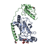
| |||||||||||||||
|---|---|---|---|---|---|---|---|---|---|---|---|---|---|---|---|---|
| 1 |
| |||||||||||||||
| Unit cell |
| |||||||||||||||
| Components on special symmetry positions |
|
-Components
-Protein , 2 types, 2 molecules AB
| #1: Protein | Mass: 16755.227 Da / Num. of mol.: 1 Source method: isolated from a genetically manipulated source Source: (gene. exp.) Homo sapiens (human) / Gene: UBE2D2, PUBC1, UBC4, UBC5B, UBCH4, UBCH5B / Plasmid: pETDUET / Production host:  |
|---|---|
| #2: Protein | Mass: 15597.544 Da / Num. of mol.: 1 / Fragment: UNP residues 126-258 Source method: isolated from a genetically manipulated source Source: (gene. exp.) Homo sapiens (human) / Gene: RNF25 / Plasmid: pETDUET / Production host: References: UniProt: Q96BH1, Ligases; Forming carbon-nitrogen bonds; Acid-amino-acid ligases (peptide synthases) |
-Non-polymers , 5 types, 259 molecules 


| #3: Chemical |  Mass: 62.068 Da / Num. of mol.: 3 / Source method: obtained synthetically / Formula: C2H6O2 Mass: 62.068 Da / Num. of mol.: 3 / Source method: obtained synthetically / Formula: C2H6O2#4: Chemical | ChemComp-PEG / |  Mass: 106.120 Da / Num. of mol.: 1 / Source method: obtained synthetically / Formula: C4H10O3 Mass: 106.120 Da / Num. of mol.: 1 / Source method: obtained synthetically / Formula: C4H10O3#5: Chemical | ChemComp-OXL / |  Mass: 88.019 Da / Num. of mol.: 1 / Source method: obtained synthetically / Formula: C2O4 Mass: 88.019 Da / Num. of mol.: 1 / Source method: obtained synthetically / Formula: C2O4#6: Chemical |  Mass: 65.409 Da / Num. of mol.: 2 / Source method: obtained synthetically / Formula: Zn Mass: 65.409 Da / Num. of mol.: 2 / Source method: obtained synthetically / Formula: Zn#7: Water | ChemComp-HOH / | Mass: 18.015 Da / Num. of mol.: 252 / Source method: isolated from a natural source / Formula: H2O |
|---|
-Experimental details
-Experiment
| Experiment | Method: X-RAY DIFFRACTION / Number of used crystals: 1 |
|---|
- Sample preparation
| Crystal | Density Matthews: 2.23 Å3/Da / Density % sol: 44.86 % |
|---|---|
| Crystal grow | Temperature: 292 K / Method: vapor diffusion, hanging drop / pH: 7.5 Details: 20% PEG-10000, 8% ethylene glycol, 0.1M HEPES, pH 7.5, VAPOR DIFFUSION, HANGING DROP, temperature 292K |
-Data collection
| Diffraction | Mean temperature: 100 K | |||||||||||||||||||||||||||||||||||||||||||||||||||||||||||||||||||||||||||||
|---|---|---|---|---|---|---|---|---|---|---|---|---|---|---|---|---|---|---|---|---|---|---|---|---|---|---|---|---|---|---|---|---|---|---|---|---|---|---|---|---|---|---|---|---|---|---|---|---|---|---|---|---|---|---|---|---|---|---|---|---|---|---|---|---|---|---|---|---|---|---|---|---|---|---|---|---|---|---|
| Diffraction source | Source: ROTATING ANODE / Type: RIGAKU MICROMAX-007 HF / Wavelength: 1.5418 Å | |||||||||||||||||||||||||||||||||||||||||||||||||||||||||||||||||||||||||||||
| Detector | Type: MAR scanner 345 mm plate / Detector: IMAGE PLATE / Date: Nov 18, 2009 / Details: mirrors | |||||||||||||||||||||||||||||||||||||||||||||||||||||||||||||||||||||||||||||
| Radiation | Monochromator: MIRRORS / Protocol: SINGLE WAVELENGTH / Monochromatic (M) / Laue (L): M / Scattering type: x-ray | |||||||||||||||||||||||||||||||||||||||||||||||||||||||||||||||||||||||||||||
| Radiation wavelength | Wavelength: 1.5418 Å / Relative weight: 1 | |||||||||||||||||||||||||||||||||||||||||||||||||||||||||||||||||||||||||||||
| Reflection | Resolution: 1.78→50 Å / Num. all: 27616 / Num. obs: 27367 / % possible obs: 99.1 % / Observed criterion σ(F): 0 / Observed criterion σ(I): 0 / Redundancy: 7 % / Biso Wilson estimate: 23.52 Å2 / Rmerge(I) obs: 0.057 / Χ2: 1.037 / Net I/σ(I): 11.8 | |||||||||||||||||||||||||||||||||||||||||||||||||||||||||||||||||||||||||||||
| Reflection shell | Diffraction-ID: 1
|
- Processing
| Software |
| ||||||||||||||||||||||||||||||||||||||||||||||||||||||||
|---|---|---|---|---|---|---|---|---|---|---|---|---|---|---|---|---|---|---|---|---|---|---|---|---|---|---|---|---|---|---|---|---|---|---|---|---|---|---|---|---|---|---|---|---|---|---|---|---|---|---|---|---|---|---|---|---|---|
| Refinement | Method to determine structure: MOLECULAR REPLACEMENT Starting model: 2ESK Resolution: 1.78→33.339 Å / Occupancy max: 1 / Occupancy min: 0.27 / SU ML: 0.24 / Cross valid method: THROUGHOUT / σ(F): 1.35 / Phase error: 19.76 / Stereochemistry target values: Engh & Huber
| ||||||||||||||||||||||||||||||||||||||||||||||||||||||||
| Solvent computation | Shrinkage radii: 0.9 Å / VDW probe radii: 1.11 Å / Solvent model: FLAT BULK SOLVENT MODEL / Bsol: 43.517 Å2 / ksol: 0.346 e/Å3 | ||||||||||||||||||||||||||||||||||||||||||||||||||||||||
| Displacement parameters | Biso max: 109.11 Å2 / Biso mean: 29.4482 Å2 / Biso min: 12.12 Å2
| ||||||||||||||||||||||||||||||||||||||||||||||||||||||||
| Refinement step | Cycle: LAST / Resolution: 1.78→33.339 Å
| ||||||||||||||||||||||||||||||||||||||||||||||||||||||||
| Refine LS restraints |
| ||||||||||||||||||||||||||||||||||||||||||||||||||||||||
| LS refinement shell | Refine-ID: X-RAY DIFFRACTION / Total num. of bins used: 6
|
