 Movie
Movie Controller


+ Open data

- Basic information
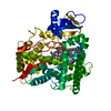
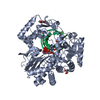
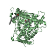
| Entry | Database: PDB / ID: 3h5y | ||||||
|---|---|---|---|---|---|---|---|
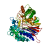
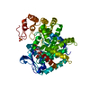
| Title | Norovirus polymerase+primer/template+CTP complex at 6 mM MnCl2 | ||||||
 Components Components |
| ||||||
 Keywords Keywords | Transferase/RNA / caliciviruses / viral RNA polymerase / Hydrolase / Nucleotide-binding / Nucleotidyltransferase / Protease / RNA replication / RNA-directed RNA polymerase / Thiol protease / Transferase / Transferase-RNA COMPLEX | ||||||
| Function / homology |  Function and homology information Function and homology informationribonucleoside triphosphate phosphatase activity / host cell / cysteine-type endopeptidase activity / viral RNA genome replication / RNA-directed RNA polymerase activity / DNA-templated transcription / proteolysis / RNA binding / ATP binding / metal ion binding Similarity search - Function | ||||||
| Biological species |   Norwalk virus Norwalk virus | ||||||
| Method |  X-RAY DIFFRACTION / SYNCHROTRON / FOURIER SYNTHESIS / Resolution: 1.77 Å X-RAY DIFFRACTION / SYNCHROTRON / FOURIER SYNTHESIS / Resolution: 1.77 Å | ||||||
 Authors Authors | Zamyatkin, D.F. / Parra, F. / Machin, A. / Grochulski, P. / Ng, K.K.S. | ||||||
 Citation Citation | Journal: J.Mol.Biol. / Year: 2009 Title: Binding of 2'-amino-2'-deoxycytidine-5'-triphosphate to norovirus polymerase induces rearrangement of the active site. Authors: Zamyatkin, D.F. / Parra, F. / Machin, A. / Grochulski, P. / Ng, K.K. | ||||||
| History |
|
- Structure visualization
| Structure viewer | Molecule: MolmilJmol/JSmol |
|---|
- Downloads & links
-Download
| PDBx/mmCIF format | 3h5y.cif.gz | 129.6 KB | Display | PDBx/mmCIF format |
|---|---|---|---|---|
| PDB format | pdb3h5y.ent.gz | 95.6 KB | Display | PDB format |
| PDBx/mmJSON format | 3h5y.json.gz | Tree view | PDBx/mmJSON format | |
| Others |  Other downloads Other downloads |
-Validation report
| Arichive directory | https://data.pdbj.org/pub/pdb/validation_reports/h5/3h5yftp://data.pdbj.org/pub/pdb/validation_reports/h5/3h5y | HTTPS FTP |
|---|
-Related structure data
| Related structure data |  3h5xC  3bsoS S: Starting model for refinement C: citing same article ( |
|---|---|
| Similar structure data |


-Links
 PDBj
PDBj
- Assembly
| Deposited unit | 
| ||||||||
|---|---|---|---|---|---|---|---|---|---|
| 1 |
| ||||||||
| Unit cell |
|
-Components
-Protein , 1 types, 1 molecules A
| #1: Protein | Mass: 56843.574 Da / Num. of mol.: 1 Source method: isolated from a genetically manipulated source Source: (gene. exp.) Norwalk virus / Genus: Norovirus / Strain: AST6139/01/SP / Gene: polymerase / Plasmid: pGEX-2T / Production host:  |
|---|
-RNA chain , 2 types, 2 molecules PT
| #2: RNA chain | Mass: 2557.577 Da / Num. of mol.: 1 / Source method: obtained synthetically Details: Chemically synthesized self-complementary RNA oligonucleotide with 2-base overhang |
|---|---|
| #3: RNA chain | Mass: 2862.759 Da / Num. of mol.: 1 / Source method: obtained synthetically Details: Chemically synthesized self-complementary RNA oligonucleotide that was extended by a single cytidine-5'-monophosphate residue during crystallization |
-Non-polymers , 4 types, 381 molecules 


| #4: Chemical | ChemComp-MN /  Mass: 54.938 Da / Num. of mol.: 4 / Source method: obtained synthetically / Formula: Mn Mass: 54.938 Da / Num. of mol.: 4 / Source method: obtained synthetically / Formula: Mn#5: Chemical | ChemComp-CTP / |  Mass: 483.156 Da / Num. of mol.: 1 / Source method: obtained synthetically / Formula: C9H16N3O14P3 Mass: 483.156 Da / Num. of mol.: 1 / Source method: obtained synthetically / Formula: C9H16N3O14P3#6: Chemical | ChemComp-GOL /  Mass: 92.094 Da / Num. of mol.: 4 / Source method: obtained synthetically / Formula: C3H8O3 Mass: 92.094 Da / Num. of mol.: 4 / Source method: obtained synthetically / Formula: C3H8O3#7: Water | ChemComp-HOH / | Mass: 18.015 Da / Num. of mol.: 372 / Source method: isolated from a natural source / Formula: H2O |
|---|
-Experimental details
-Experiment
| Experiment | Method: X-RAY DIFFRACTION / Number of used crystals: 1 |
|---|
- Sample preparation
| Crystal | Density Matthews: 2.69 Å3/Da / Density % sol: 54.2 % | ||||||||||||||||||||||||||||||||||||||||||||||||||||||||||||||||||||
|---|---|---|---|---|---|---|---|---|---|---|---|---|---|---|---|---|---|---|---|---|---|---|---|---|---|---|---|---|---|---|---|---|---|---|---|---|---|---|---|---|---|---|---|---|---|---|---|---|---|---|---|---|---|---|---|---|---|---|---|---|---|---|---|---|---|---|---|---|---|
| Crystal grow | Temperature: 298 K / Method: vapor diffusion, hanging drop / pH: 7 Details: 160 g/L PEG 8000, 250 g/L glycerol, 100 mM Tris-Cl, 50 mM KCl, 4 mM MgCl2, 6 mM MnCl2, 14 mM mercaptoethanol, 1 g/L CHAPS, pH 7.0, VAPOR DIFFUSION, HANGING DROP, temperature 298K | ||||||||||||||||||||||||||||||||||||||||||||||||||||||||||||||||||||
| Components of the solutions |
|
-Data collection
| Diffraction | Mean temperature: 100 K |
|---|---|
| Diffraction source | Source: SYNCHROTRON / Site: CLSI  / Beamline: 08ID-1 / Wavelength: 0.97934 Å / Beamline: 08ID-1 / Wavelength: 0.97934 Å |
| Detector | Type: MARMOSAIC 225 mm CCD / Detector: CCD / Date: Feb 25, 2008 Details: WHITE BEAM SLITS, DOUBLE CRYSTAL MONOCHROMATOR (DCM), VERTICALLY FOCUSING MIRROR (VFM) |
| Radiation | Monochromator: CRYO-COOLED FIRST AND SAGITTALLY BENT SECOND CRYSTAL OF DOUBLE CRYSTAL MONOCHROMATOR Protocol: SINGLE WAVELENGTH / Monochromatic (M) / Laue (L): M / Scattering type: x-ray |
| Radiation wavelength | Wavelength: 0.97934 Å / Relative weight: 1 |
| Reflection | Resolution: 1.77→20 Å / Num. all: 65904 / Num. obs: 65904 / % possible obs: 99.9 % / Observed criterion σ(F): 0 / Observed criterion σ(I): 0 / Redundancy: 6.1 % / Biso Wilson estimate: 36.9 Å2 / Rmerge(I) obs: 0.044 / Rsym value: 0.044 / Net I/σ(I): 23.1 |
| Reflection shell | Resolution: 1.77→1.97 Å / Redundancy: 6.1 % / Rmerge(I) obs: 0.622 / Mean I/σ(I) obs: 3.2 / Num. unique all: 17876 / Rsym value: 0.622 / % possible all: 100 |
- Processing
| Software |
| |||||||||||||||||||||||||||||||||||||||||||||||||||||||||||||||||
|---|---|---|---|---|---|---|---|---|---|---|---|---|---|---|---|---|---|---|---|---|---|---|---|---|---|---|---|---|---|---|---|---|---|---|---|---|---|---|---|---|---|---|---|---|---|---|---|---|---|---|---|---|---|---|---|---|---|---|---|---|---|---|---|---|---|---|
| Refinement | Method to determine structure: FOURIER SYNTHESIS Starting model: 3BSO Resolution: 1.77→19.74 Å / Cor.coef. Fo:Fc: 0.954 / Cor.coef. Fo:Fc free: 0.94 / SU B: 2.616 / SU ML: 0.083 / Isotropic thermal model: Isotropic / Cross valid method: THROUGHOUT / σ(F): 0 / σ(I): 0 / ESU R: 0.117 / ESU R Free: 0.115 / Stereochemistry target values: MAXIMUM LIKELIHOOD
| |||||||||||||||||||||||||||||||||||||||||||||||||||||||||||||||||
| Solvent computation | Ion probe radii: 0.8 Å / Shrinkage radii: 0.8 Å / VDW probe radii: 1.4 Å / Solvent model: BABINET MODEL WITH MASK | |||||||||||||||||||||||||||||||||||||||||||||||||||||||||||||||||
| Displacement parameters | Biso mean: 34.253 Å2
| |||||||||||||||||||||||||||||||||||||||||||||||||||||||||||||||||
| Refinement step | Cycle: LAST / Resolution: 1.77→19.74 Å
| |||||||||||||||||||||||||||||||||||||||||||||||||||||||||||||||||
| Refine LS restraints |
| |||||||||||||||||||||||||||||||||||||||||||||||||||||||||||||||||
| LS refinement shell | Resolution: 1.77→1.816 Å / Total num. of bins used: 20
|
