 Movie
Movie Controller

[English] 日本語
 Yorodumi
- PDB-3h5t: Crystal structure of a transcriptional regulator, Lacl family pro... -
+ Open data

- Basic information
| Entry | Database: PDB / ID: 3h5t | ||||||
|---|---|---|---|---|---|---|---|
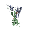
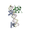
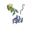
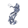
| Title | Crystal structure of a transcriptional regulator, Lacl family protein from Corynebacterium glutamicum | ||||||
 Components Components | Transcriptional regulator, LacI family | ||||||
 Keywords Keywords | transcription regulator / Transcriptional regulator / DNA-dependent / Protein Structure Initiative II(PSI II) / NYSGXRC / 11232d) / Structural Genomics / New York SGX Research Center for Structural Genomics / DNA-binding / Transcription / Transcription regulation | ||||||
| Function / homology | lambda repressor-like DNA-binding domains / 434 Repressor (Amino-terminal Domain) / Response regulator / Rossmann fold / Orthogonal Bundle / 3-Layer(aba) Sandwich / Mainly Alpha / Alpha Beta / :  Function and homology information Function and homology information | ||||||
| Biological species |  Corynebacterium glutamicum (bacteria) Corynebacterium glutamicum (bacteria) | ||||||
| Method |  X-RAY DIFFRACTION / SYNCHROTRON / SAD / Resolution: 2.53 Å X-RAY DIFFRACTION / SYNCHROTRON / SAD / Resolution: 2.53 Å | ||||||
 Authors Authors | Palani, K. / Burley, S.K. / Swaminathan, S. / New York SGX Research Center for Structural Genomics (NYSGXRC) | ||||||
 Citation Citation | Journal: To be Published Title: Crystal structure of a transcriptional regulator, Lacl family protein from Corynebacterium glutamicum Authors: Palani, K. / Burley, S.K. / Swaminathan, S. | ||||||
| History |
|
- Structure visualization
| Structure viewer | Molecule: MolmilJmol/JSmol |
|---|
- Downloads & links
-Download
| PDBx/mmCIF format | 3h5t.cif.gz | 77.6 KB | Display | PDBx/mmCIF format |
|---|---|---|---|---|
| PDB format | pdb3h5t.ent.gz | 58.1 KB | Display | PDB format |
| PDBx/mmJSON format | 3h5t.json.gz | Tree view | PDBx/mmJSON format | |
| Others |  Other downloads Other downloads |
-Validation report
| Summary document | 3h5t_validation.pdf.gz | 427.2 KB | Display | wwPDB validaton report |
|---|---|---|---|---|
| Full document | 3h5t_full_validation.pdf.gz | 433.1 KB | Display | |
| Data in XML | 3h5t_validation.xml.gz | 15.2 KB | Display | |
| Data in CIF | 3h5t_validation.cif.gz | 21 KB | Display | |
| Arichive directory | https://data.pdbj.org/pub/pdb/validation_reports/h5/3h5tftp://data.pdbj.org/pub/pdb/validation_reports/h5/3h5t | HTTPS FTP |
-Related structure data
| Similar structure data | |
|---|---|
| Other databases |


-Links
 PDBj
PDBj- Assembly
| Deposited unit | 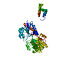
| ||||||||
|---|---|---|---|---|---|---|---|---|---|
| 1 |
| ||||||||
| 2 | 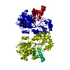
| ||||||||
| Unit cell |
|
-Components
| #1: Protein | Mass: 39664.777 Da / Num. of mol.: 1 Source method: isolated from a genetically manipulated source Source: (gene. exp.) Corynebacterium glutamicum (bacteria) / Gene: cg2910 / Plasmid: BC-pSGX3 (BC) / Production host: |
|---|---|
| #2: Water | ChemComp-HOH /  Mass: 18.015 Da / Num. of mol.: 109 / Source method: isolated from a natural source / Formula: H2O Mass: 18.015 Da / Num. of mol.: 109 / Source method: isolated from a natural source / Formula: H2O |
-Experimental details
-Experiment
| Experiment | Method: X-RAY DIFFRACTION / Number of used crystals: 1 |
|---|
- Sample preparation
| Crystal grow | Temperature: 298 K / Method: vapor diffusion, sitting drop / pH: 7 Details: 0.2M Lithium acetate dihydrate, 20% Polyethylene glycol 3350, pH 7.0, VAPOR DIFFUSION, SITTING DROP, temperature 298.0K |
|---|
-Data collection
| Diffraction | Mean temperature: 100 K |
|---|---|
| Diffraction source | Source: SYNCHROTRON / Site: NSLS  / Beamline: X25 / Wavelength: 0.979 Å / Beamline: X25 / Wavelength: 0.979 Å |
| Detector | Type: ADSC QUANTUM 315 / Detector: CCD / Date: Jan 28, 2009 / Details: MIRRORS |
| Radiation | Monochromator: Si(III) / Protocol: SINGLE WAVELENGTH / Monochromatic (M) / Laue (L): M / Scattering type: x-ray |
| Radiation wavelength | Wavelength: 0.979 Å / Relative weight: 1 |
| Reflection | Resolution: 2.53→47.21 Å / Num. all: 27874 / Num. obs: 27874 / % possible obs: 96.2 % / Observed criterion σ(F): 0 / Observed criterion σ(I): 0 / Redundancy: 6.9 % / Biso Wilson estimate: 22.3 Å2 / Rmerge(I) obs: 0.081 / Net I/σ(I): 20.6 |
| Reflection shell | Resolution: 2.53→2.62 Å / Redundancy: 4.4 % / Rmerge(I) obs: 0.294 / Mean I/σ(I) obs: 2.5 / Num. unique all: 2222 / % possible all: 78.2 |
- Processing
| Software |
| |||||||||||||||||||||||||
|---|---|---|---|---|---|---|---|---|---|---|---|---|---|---|---|---|---|---|---|---|---|---|---|---|---|---|
| Refinement | Method to determine structure: SAD / Resolution: 2.53→47.21 Å / Rfactor Rfree error: 0.008 / Data cutoff high absF: 168737.88 / Data cutoff low absF: 0 / Isotropic thermal model: RESTRAINED / Cross valid method: THROUGHOUT / σ(F): 0 / Stereochemistry target values: Engh & Huber
| |||||||||||||||||||||||||
| Solvent computation | Solvent model: FLAT MODEL / Bsol: 28.2459 Å2 / ksol: 0.350177 e/Å3 | |||||||||||||||||||||||||
| Displacement parameters | Biso mean: 30 Å2
| |||||||||||||||||||||||||
| Refine analyze |
| |||||||||||||||||||||||||
| Refinement step | Cycle: LAST / Resolution: 2.53→47.21 Å
| |||||||||||||||||||||||||
| Refine LS restraints |
| |||||||||||||||||||||||||
| LS refinement shell | Resolution: 2.53→2.69 Å / Rfactor Rfree error: 0.023 / Total num. of bins used: 6
| |||||||||||||||||||||||||
| Xplor file |
|
