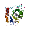
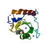
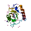
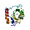
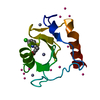
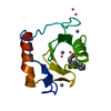
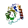
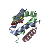
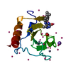
[English] 日本語
Yorodumi
- PDB-3gv4: Crystal structure of human HDAC6 zinc finger domain and ubiquitin... -

+
Open data


ID or keywords:

Loading...

-
Basic information

Entry
Database: PDB / ID: 3gv4
TitleCrystal structure of human HDAC6 zinc finger domain and ubiquitin C-terminal peptide RLRGG
Components
  • Histone deacetylase 6
  • ubiquitin C-terminal peptide RLRGG
KeywordsHYDROLASE / HDAC6 / zinc finger / ubiquitin C-terminal peptide RLRGG / SGC / Actin-binding / Chromatin regulator / Cytoplasm / Metal-binding / Nucleus / Phosphoprotein / Polymorphism / Repressor / Transcription / Transcription regulation / Ubl conjugation / Zinc / Zinc-finger / Structural Genomics / Structural Genomics Consortium
Function / homology
Function and homology information


negative regulation of hydrogen peroxide metabolic process / cellular response to topologically incorrect protein / polyubiquitinated misfolded protein transport / positive regulation of cellular response to oxidative stress / erythrocyte enucleation / negative regulation of aggrephagy / positive regulation of cholangiocyte proliferation / response to misfolded protein / positive regulation of protein oligomerization / negative regulation of axon extension involved in axon guidance ...negative regulation of hydrogen peroxide metabolic process / cellular response to topologically incorrect protein / polyubiquitinated misfolded protein transport / positive regulation of cellular response to oxidative stress / erythrocyte enucleation / negative regulation of aggrephagy / positive regulation of cholangiocyte proliferation / response to misfolded protein / positive regulation of protein oligomerization / negative regulation of axon extension involved in axon guidance / regulation of microtubule-based movement / type 2 mitophagy / negative regulation of protein-containing complex disassembly / peroxidase inhibitor activity / regulation of establishment of protein localization / regulation of autophagy of mitochondrion / protein-containing complex disassembly / tubulin deacetylation / Cilium Assembly / collateral sprouting / tubulin deacetylase activity / Transcriptional regulation by RUNX2 / lysosome localization / negative regulation of microtubule depolymerization / positive regulation of dendrite morphogenesis / ATPase inhibitor activity / cilium disassembly / misfolded protein binding / ubiquitin-dependent protein catabolic process via the multivesicular body sorting pathway / positive regulation of type 2 mitophagy / histone H4K16 deacetylase activity, hydrolytic mechanism / histone H4K5 deacetylase activity, hydrolytic mechanism / histone H4K8 deacetylase activity, hydrolytic mechanism / histone H3K4 deacetylase activity, hydrolytic mechanism / histone H3K14 deacetylase activity, hydrolytic mechanism / dendritic spine morphogenesis / histone H4K12 deacetylase activity, hydrolytic mechanism / protein deacetylation / histone H3K9 deacetylase activity, hydrolytic mechanism / aggresome assembly / regulation of androgen receptor signaling pathway / regulation of mitochondrion organization / cellular response to misfolded protein / Transferases; Acyltransferases; Aminoacyltransferases / regulation of fat cell differentiation / protein lysine deacetylase activity / microtubule associated complex / Hydrolases; Acting on carbon-nitrogen bonds, other than peptide bonds; In linear amides / aggresome / cellular response to parathyroid hormone stimulus / response to corticosterone / histone deacetylase activity / positive regulation of intracellular estrogen receptor signaling pathway / Notch-HLH transcription pathway / negative regulation of gene expression, epigenetic / axonal transport of mitochondrion / dynein complex binding / beta-tubulin binding / protein quality control for misfolded or incompletely synthesized proteins / RUNX2 regulates osteoblast differentiation / cell leading edge / response to dexamethasone / histone deacetylase complex / response to immobilization stress / positive regulation of epithelial cell migration / cilium assembly / polyubiquitin modification-dependent protein binding / alpha-tubulin binding / regulation of macroautophagy / HSF1 activation / negative regulation of protein-containing complex assembly / inclusion body / negative regulation of proteolysis / axon cytoplasm / multivesicular body / response to amphetamine / positive regulation of synaptic transmission, glutamatergic / epigenetic regulation of gene expression / ubiquitin binding / actin filament organization / transcription corepressor binding / intracellular protein transport / Hsp90 protein binding / Late endosomal microautophagy / protein destabilization / regulation of protein stability / beta-catenin binding / caveola / NOTCH1 Intracellular Domain Regulates Transcription / tau protein binding / cellular response to hydrogen peroxide / Constitutive Signaling by NOTCH1 PEST Domain Mutants / Constitutive Signaling by NOTCH1 HD+PEST Domain Mutants / epidermal growth factor receptor signaling pathway / histone deacetylase binding / Chaperone Mediated Autophagy / protein polyubiquitination / Aggrephagy / actin binding / cellular response to heat
Similarity search - Function
Ubiquitin Carboxyl-terminal Hydrolase-like zinc finger / Zinc finger, UBP-type / Zn-finger in ubiquitin-hydrolases and other protein / Zinc finger UBP-type profile. / : / Histone deacetylase family / Histone deacetylase domain / Histone deacetylase domain superfamily / Histone deacetylase domain / Zinc/RING finger domain, C3HC4 (zinc finger) ...Ubiquitin Carboxyl-terminal Hydrolase-like zinc finger / Zinc finger, UBP-type / Zn-finger in ubiquitin-hydrolases and other protein / Zinc finger UBP-type profile. / : / Histone deacetylase family / Histone deacetylase domain / Histone deacetylase domain superfamily / Histone deacetylase domain / Zinc/RING finger domain, C3HC4 (zinc finger) / Herpes Virus-1 / Ureohydrolase domain superfamily / Zinc finger, RING/FYVE/PHD-type / 2-Layer Sandwich / Alpha Beta
Similarity search - Domain/homology
Protein deacetylase HDAC6
Similarity search - Component
Biological speciesHomo sapiens (human)
MethodX-RAY DIFFRACTION / MOLECULAR REPLACEMENT / Resolution: 1.72 Å
AuthorsDong, A. / Ravichandran, M. / Loppnau, P. / Li, Y. / MacKenzie, F. / Kozieradzki, I. / Edwards, A.M. / Arrowsmith, C.H. / Weigelt, J. / Bountra, C. ...Dong, A. / Ravichandran, M. / Loppnau, P. / Li, Y. / MacKenzie, F. / Kozieradzki, I. / Edwards, A.M. / Arrowsmith, C.H. / Weigelt, J. / Bountra, C. / Bochkarev, A. / Dhe-Paganon, S. / Min, J. / Ouyang, H. / Structural Genomics Consortium (SGC)
CitationJournal: To be Published
Title: Crystal structure of human HDAC6 zinc finger domain and ubiquitin C-terminal peptide RLRGG
Authors: Dong, A. / Ravichandran, M. / Loppnau, P. / Li, Y. / MacKenzie, F. / Kozieradzki, I. / Edwards, A.M. / Arrowsmith, C.H. / Weigelt, J. / Bountra, C. / Bochkarev, A. / Dhe-Paganon, S. / Min, J. / Ouyang, H.
History
DepositionMar 30, 2009Deposition site: RCSB / Processing site: RCSB
Revision 1.0Apr 28, 2009Provider: repository / Type: Initial release
Revision 1.1Jul 13, 2011Group: Version format compliance
Revision 1.2Nov 1, 2017Group: Refinement description / Category: software / Item: _software.name
Revision 1.3Sep 6, 2023Group: Data collection / Database references ...Data collection / Database references / Derived calculations / Refinement description
Category: chem_comp_atom / chem_comp_bond ...chem_comp_atom / chem_comp_bond / database_2 / pdbx_initial_refinement_model / struct_site
Item: _database_2.pdbx_DOI / _database_2.pdbx_database_accession ..._database_2.pdbx_DOI / _database_2.pdbx_database_accession / _struct_site.pdbx_auth_asym_id / _struct_site.pdbx_auth_comp_id / _struct_site.pdbx_auth_seq_id

-
Structure visualization

Structure viewerMolecule:
MolmilJmol/JSmol

Downloads & links

-
Assembly

Deposited unit
A: Histone deacetylase 6
H: ubiquitin C-terminal peptide RLRGG
hetero molecules


Theoretical massNumber of molelcules
Total (without water)12,8206
Polymers12,5832
Non-polymers2364
Water3,693205
1


  • Idetical with deposited unit
  • defined by author&software
TypeNameSymmetry operationNumber
identity operation1_555x,y,z1
Buried area680 Å2
ΔGint-6 kcal/mol
Surface area5500 Å2
MethodPISA
Unit cell
Length a, b, c (Å)31.657, 36.093, 88.998
Angle α, β, γ (deg.)90.00, 90.00, 90.00
Int Tables number19
Space group name H-MP212121

-
Components

#1: Protein Histone deacetylase 6 / HD6


Mass: 12023.739 Da / Num. of mol.: 1 / Fragment: UNP residues 1109-1215
Source method: isolated from a genetically manipulated source
Source: (gene. exp.) Homo sapiens (human) / Gene: HDAC6, KIAA0901, JM21 / Plasmid: P28A-LIC / Production host: Escherichia coli (E. coli) / Strain (production host): BL21(DE3) / References: UniProt: Q9UBN7, histone deacetylase
#2: Protein/peptide ubiquitin C-terminal peptide RLRGG


Mass: 559.664 Da / Num. of mol.: 1 / Source method: obtained synthetically / Details: ubiquitin C-terminal peptide
#3: Chemical ChemComp-ZN / ZINC ION


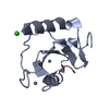
Mass: 65.409 Da / Num. of mol.: 3 / Source method: obtained synthetically / Formula: Zn
#4: Chemical ChemComp-CA / CALCIUM ION


Mass: 40.078 Da / Num. of mol.: 1 / Source method: obtained synthetically / Formula: Ca
#5: Water ChemComp-HOH / water


Mass: 18.015 Da / Num. of mol.: 205 / Source method: isolated from a natural source / Formula: H2O

-
Experimental details

-
Experiment

ExperimentMethod: X-RAY DIFFRACTION / Number of used crystals: 1

-
Sample preparation

CrystalDensity Matthews: 2.02 Å3/Da / Density % sol: 39.12 %
Crystal growTemperature: 297 K / Method: vapor diffusion, sitting drop
Details: 20% PEG3350, 0.2 M NaCl, VAPOR DIFFUSION, SITTING DROP, temperature 297K

-
Data collection

DiffractionMean temperature: 100 K
Diffraction sourceSource: ROTATING ANODE / Type: RIGAKU FR-E SUPERBRIGHT / Wavelength: 1.54 Å
DetectorType: RIGAKU RAXIS IV++ / Detector: IMAGE PLATE / Date: Apr 14, 2008 / Details: VariMax HR
RadiationProtocol: SINGLE WAVELENGTH / Monochromatic (M) / Laue (L): M / Scattering type: x-ray
Radiation wavelengthWavelength: 1.54 Å / Relative weight: 1
ReflectionResolution: 1.72→50 Å / Num. all: 10865 / Num. obs: 10865 / % possible obs: 94.5 % / Observed criterion σ(F): 0 / Observed criterion σ(I): 0 / Redundancy: 5.8 % / Biso Wilson estimate: 22.9 Å2 / Rmerge(I) obs: 0.073 / Rsym value: 0.073 / Net I/σ(I): 14.1
Reflection shellResolution: 1.72→1.78 Å / Redundancy: 3.1 % / Rmerge(I) obs: 0.231 / Mean I/σ(I) obs: 4.26 / Num. unique all: 813 / Rsym value: 0.231 / % possible all: 72.5

-
Processing

Software
NameVersionClassification
SBC-Collectdata collection
PHASESphasing
REFMAC5.2.0019refinement
Coot0.3.3model building
HKL-2000data reduction
HKL-2000data scaling
RefinementMethod to determine structure: MOLECULAR REPLACEMENT
Starting model: PDB entry 3C5K
Resolution: 1.72→33.45 Å / Cor.coef. Fo:Fc: 0.961 / Cor.coef. Fo:Fc free: 0.938 / SU B: 2.51 / SU ML: 0.082 / Cross valid method: THROUGHOUT / σ(F): 0 / σ(I): 0 / ESU R: 0.125 / ESU R Free: 0.129 / Stereochemistry target values: MAXIMUM LIKELIHOOD / Details: HYDROGENS HAVE BEEN ADDED IN THE RIDING POSITIONS
RfactorNum. reflection% reflectionSelection details
Rfree0.23034 609 5.6 %RANDOM
Rwork0.17382 ---
all0.17695 10213 --
obs0.17695 10213 94.64 %-
Solvent computationIon probe radii: 0.8 Å / Shrinkage radii: 0.8 Å / VDW probe radii: 1.4 Å / Solvent model: MASK
Displacement parametersBiso mean: 21.958 Å2
Baniso -1Baniso -2Baniso -3
1-0.01 Å20 Å20 Å2
2---1.31 Å20 Å2
3---1.3 Å2
Refinement stepCycle: LAST / Resolution: 1.72→33.45 Å
ProteinNucleic acidLigandSolventTotal
Num. atoms799 0 4 205 1008
Refine LS restraints
Refine-IDTypeDev idealDev ideal targetNumber
X-RAY DIFFRACTIONr_bond_refined_d0.010.021825
X-RAY DIFFRACTIONr_bond_other_d
X-RAY DIFFRACTIONr_angle_refined_deg1.1911.9211128
X-RAY DIFFRACTIONr_angle_other_deg
X-RAY DIFFRACTIONr_dihedral_angle_1_deg5.8135101
X-RAY DIFFRACTIONr_dihedral_angle_2_deg36.52723.88936
X-RAY DIFFRACTIONr_dihedral_angle_3_deg11.32215116
X-RAY DIFFRACTIONr_dihedral_angle_4_deg16.224152
X-RAY DIFFRACTIONr_chiral_restr0.0930.2120
X-RAY DIFFRACTIONr_gen_planes_refined0.0050.02640
X-RAY DIFFRACTIONr_gen_planes_other
X-RAY DIFFRACTIONr_nbd_refined0.1930.2438
X-RAY DIFFRACTIONr_nbd_other
X-RAY DIFFRACTIONr_nbtor_refined0.3030.2551
X-RAY DIFFRACTIONr_nbtor_other
X-RAY DIFFRACTIONr_xyhbond_nbd_refined0.1530.2121
X-RAY DIFFRACTIONr_xyhbond_nbd_other
X-RAY DIFFRACTIONr_metal_ion_refined0.0370.23
X-RAY DIFFRACTIONr_metal_ion_other
X-RAY DIFFRACTIONr_symmetry_vdw_refined0.2080.233
X-RAY DIFFRACTIONr_symmetry_vdw_other
X-RAY DIFFRACTIONr_symmetry_hbond_refined0.1620.226
X-RAY DIFFRACTIONr_symmetry_hbond_other
X-RAY DIFFRACTIONr_symmetry_metal_ion_refined0.0390.21
X-RAY DIFFRACTIONr_symmetry_metal_ion_other
X-RAY DIFFRACTIONr_mcbond_it0.6121.5520
X-RAY DIFFRACTIONr_mcbond_other
X-RAY DIFFRACTIONr_mcangle_it1.022821
X-RAY DIFFRACTIONr_scbond_it1.5953355
X-RAY DIFFRACTIONr_scangle_it2.474.5307
X-RAY DIFFRACTIONr_rigid_bond_restr
X-RAY DIFFRACTIONr_sphericity_free
X-RAY DIFFRACTIONr_sphericity_bonded
LS refinement shellResolution: 1.72→1.762 Å / Total num. of bins used: 20
RfactorNum. reflection% reflection
Rfree0.477 34 -
Rwork0.378 523 -
obs--67.68 %

+
About Yorodumi

-
News

-
Feb 9, 2022. New format data for meta-information of EMDB entries

New format data for meta-information of EMDB entries

  • Version 3 of the EMDB header file is now the official format.
  • The previous official version 1.9 will be removed from the archive.

Related info.:EMDB header

External links:wwPDB to switch to version 3 of the EMDB data model

-
Aug 12, 2020. Covid-19 info

Covid-19 info

URL: https://pdbj.org/emnavi/covid19.php

New page: Covid-19 featured information page in EM Navigator.

Related info.:Covid-19 info / Mar 5, 2020. Novel coronavirus structure data

+
Mar 5, 2020. Novel coronavirus structure data

Novel coronavirus structure data

Related info.:Yorodumi Speices / Aug 12, 2020. Covid-19 info

External links:COVID-19 featured content - PDBj / Molecule of the Month (242):Coronavirus Proteases

+
Jan 31, 2019. EMDB accession codes are about to change! (news from PDBe EMDB page)

EMDB accession codes are about to change! (news from PDBe EMDB page)

  • The allocation of 4 digits for EMDB accession codes will soon come to an end. Whilst these codes will remain in use, new EMDB accession codes will include an additional digit and will expand incrementally as the available range of codes is exhausted. The current 4-digit format prefixed with “EMD-” (i.e. EMD-XXXX) will advance to a 5-digit format (i.e. EMD-XXXXX), and so on. It is currently estimated that the 4-digit codes will be depleted around Spring 2019, at which point the 5-digit format will come into force.
  • The EM Navigator/Yorodumi systems omit the EMD- prefix.

Related info.:Q: What is EMD? / ID/Accession-code notation in Yorodumi/EM Navigator

External links:EMDB Accession Codes are Changing Soon! / Contact to PDBj

+
Jul 12, 2017. Major update of PDB

Major update of PDB

  • wwPDB released updated PDB data conforming to the new PDBx/mmCIF dictionary.
  • This is a major update changing the version number from 4 to 5, and with Remediation, in which all the entries are updated.
  • In this update, many items about electron microscopy experimental information are reorganized (e.g. em_software).
  • Now, EM Navigator and Yorodumi are based on the updated data.

External links:wwPDB Remediation / Enriched Model Files Conforming to OneDep Data Standards Now Available in the PDB FTP Archive

-
Yorodumi

Thousand views of thousand structures

  • Yorodumi is a browser for structure data from EMDB, PDB, SASBDB, etc.
  • This page is also the successor to EM Navigator detail page, and also detail information page/front-end page for Omokage search.
  • The word "yorodu" (or yorozu) is an old Japanese word meaning "ten thousand". "mi" (miru) is to see.

Related info.:EMDB / PDB / SASBDB / Comparison of 3 databanks / Yorodumi Search / Aug 31, 2016. New EM Navigator & Yorodumi / Yorodumi Papers / Jmol/JSmol / Function and homology information / Changes in new EM Navigator and Yorodumi

Read more