 Movie
Movie Controller


+ Open data

- Basic information
| Entry | Database: PDB / ID: 3gt6 | ||||||
|---|---|---|---|---|---|---|---|
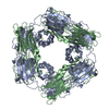
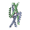
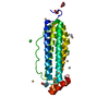
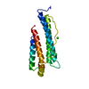
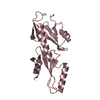
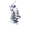
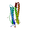
| Title | Crystal Structure of the hspA from Xanthomonas axonopodis | ||||||
 Components Components | Low molecular weight heat shock protein | ||||||
 Keywords Keywords | CHAPERONE / hspA / shp / shsp / Xanthomonas axonopodis / high resolution / stress response | ||||||
| Function / homology |  Function and homology information Function and homology information: / Immunoglobulin-like - #790 / Hsp20/alpha crystallin family / Small heat shock protein (sHSP) domain profile. / Alpha crystallin/Hsp20 domain / HSP20-like chaperone / Immunoglobulin-like / Sandwich / Mainly Beta Similarity search - Domain/homology | ||||||
| Biological species |  Xanthomonas axonopodis pv. citri (bacteria) Xanthomonas axonopodis pv. citri (bacteria) | ||||||
| Method |  X-RAY DIFFRACTION / SYNCHROTRON / MOLECULAR REPLACEMENT / molecular replacement / Resolution: 2.15 Å X-RAY DIFFRACTION / SYNCHROTRON / MOLECULAR REPLACEMENT / molecular replacement / Resolution: 2.15 Å | ||||||
 Authors Authors | Hilario, E. / Medrano, F.J. / Bertolini, M.C. | ||||||
 Citation Citation | Journal: J.Mol.Biol. / Year: 2011 Title: Crystal structures of Xanthomonas small heat shock protein provide a structural basis for an active molecular chaperone oligomer. Authors: Hilario, E. / Martin, F.J. / Bertolini, M.C. / Fan, L. | ||||||
| History |
|
- Structure visualization
| Structure viewer | Molecule: MolmilJmol/JSmol |
|---|
- Downloads & links
-Download
| PDBx/mmCIF format | 3gt6.cif.gz | 55.7 KB | Display | PDBx/mmCIF format |
|---|---|---|---|---|
| PDB format | pdb3gt6.ent.gz | 40.2 KB | Display | PDB format |
| PDBx/mmJSON format | 3gt6.json.gz | Tree view | PDBx/mmJSON format | |
| Others |  Other downloads Other downloads |
-Validation report
| Arichive directory | https://data.pdbj.org/pub/pdb/validation_reports/gt/3gt6ftp://data.pdbj.org/pub/pdb/validation_reports/gt/3gt6 | HTTPS FTP |
|---|
-Related structure data
| Related structure data | 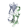 3gufC 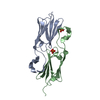 3glaS  3gtb S: Starting model for refinement C: citing same article ( |
|---|---|
| Similar structure data |


-Links
 PDBj
PDBj


- Assembly
| Deposited unit | 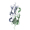
| ||||||||
|---|---|---|---|---|---|---|---|---|---|
| 1 |
| ||||||||
| Unit cell |
|
-Components
| #1: Protein | Mass: 11610.074 Da / Num. of mol.: 2 / Fragment: UNP residues 37-139 Source method: isolated from a genetically manipulated source Source: (gene. exp.) Xanthomonas axonopodis pv. citri (bacteria)Gene: hspA / Plasmid: pET28a-hspA / Production host: #2: Water | ChemComp-HOH / |  Mass: 18.015 Da / Num. of mol.: 148 / Source method: isolated from a natural source / Formula: H2O Mass: 18.015 Da / Num. of mol.: 148 / Source method: isolated from a natural source / Formula: H2O |
|---|
-Experimental details
-Experiment
| Experiment | Method: X-RAY DIFFRACTION / Number of used crystals: 1 |
|---|
- Sample preparation
| Crystal | Density Matthews: 3.79 Å3/Da / Density % sol: 67.53 % |
|---|---|
| Crystal grow | Temperature: 293 K / Method: vapor diffusion, sitting drop Details: 24% (w/v) PEG 1500, 20 % glycerol, VAPOR DIFFUSION, SITTING DROP, temperature 293 K |
-Data collection
| Diffraction | Mean temperature: 100 K |
|---|---|
| Diffraction source | Source: SYNCHROTRON / Site: LNLS  / Beamline: D03B-MX1 / Wavelength: 1.43 Å / Beamline: D03B-MX1 / Wavelength: 1.43 Å |
| Detector | Type: MAR CCD 165 mm / Detector: CCD / Date: Jan 24, 2006 |
| Radiation | Protocol: SINGLE WAVELENGTH / Monochromatic (M) / Laue (L): M / Scattering type: x-ray |
| Radiation wavelength | Wavelength: 1.43 Å / Relative weight: 1 |
| Reflection | Resolution: 2.15→26.96 Å / Num. obs: 18680 / Redundancy: 3.8 % / Rmerge(I) obs: 0.049 |
-Phasing
| Phasing | Method: molecular replacement | |||||||||
|---|---|---|---|---|---|---|---|---|---|---|
| Phasing MR | Rfactor: 28.34 / Model details: Phaser MODE: MR_AUTO
|
- Processing
| Software |
| |||||||||||||||||||||||||||||||||||||||||||||||||||||||||||||||||
|---|---|---|---|---|---|---|---|---|---|---|---|---|---|---|---|---|---|---|---|---|---|---|---|---|---|---|---|---|---|---|---|---|---|---|---|---|---|---|---|---|---|---|---|---|---|---|---|---|---|---|---|---|---|---|---|---|---|---|---|---|---|---|---|---|---|---|
| Refinement | Method to determine structure: MOLECULAR REPLACEMENT Starting model: 3GLA Resolution: 2.15→26.96 Å / Cor.coef. Fo:Fc: 0.94 / Cor.coef. Fo:Fc free: 0.917 / WRfactor Rfree: 0.234 / WRfactor Rwork: 0.196 / Occupancy max: 1 / Occupancy min: 0.5 / FOM work R set: 0.855 / SU B: 3.865 / SU ML: 0.103 / SU R Cruickshank DPI: 0.175 / SU Rfree: 0.162 / Cross valid method: THROUGHOUT / σ(F): 0 / ESU R: 0.175 / ESU R Free: 0.162 / Stereochemistry target values: MAXIMUM LIKELIHOOD Details: HYDROGENS HAVE BEEN ADDED IN THE RIDING POSITIONS U VALUES : REFINED INDIVIDUALLY
| |||||||||||||||||||||||||||||||||||||||||||||||||||||||||||||||||
| Solvent computation | Ion probe radii: 0.8 Å / Shrinkage radii: 0.8 Å / VDW probe radii: 1.4 Å / Solvent model: MASK | |||||||||||||||||||||||||||||||||||||||||||||||||||||||||||||||||
| Displacement parameters | Biso max: 60.93 Å2 / Biso mean: 26.304 Å2 / Biso min: 10.63 Å2
| |||||||||||||||||||||||||||||||||||||||||||||||||||||||||||||||||
| Refinement step | Cycle: LAST / Resolution: 2.15→26.96 Å
| |||||||||||||||||||||||||||||||||||||||||||||||||||||||||||||||||
| Refine LS restraints |
| |||||||||||||||||||||||||||||||||||||||||||||||||||||||||||||||||
| LS refinement shell | Resolution: 2.15→2.201 Å / Total num. of bins used: 20
|
