 Movie
Movie Controller

[English] 日本語
 Yorodumi
- PDB-1bg7: LOCALIZED UNFOLDING AT THE JUNCTION OF THREE FERRITIN SUBUNITS. A... -
+ Open data

- Basic information
| Entry | Database: PDB / ID: 1bg7 | ||||||
|---|---|---|---|---|---|---|---|
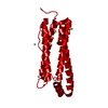
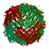
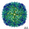
| Title | LOCALIZED UNFOLDING AT THE JUNCTION OF THREE FERRITIN SUBUNITS. A MECHANISM FOR IRON RELEASE? | ||||||
 Components Components | FERRITIN | ||||||
 Keywords Keywords | IRON STORAGE / FERRITIN | ||||||
| Function / homology |  Function and homology information Function and homology informationferroxidase / ferroxidase activity / ferric iron binding / iron ion transport / ferrous iron binding / intracellular iron ion homeostasis / cytoplasm Similarity search - Function | ||||||
| Biological species | Rana catesbeiana (American bullfrog) | ||||||
| Method |  X-RAY DIFFRACTION / SYNCHROTRON / MOLECULAR REPLACEMENT / Resolution: 1.85 Å X-RAY DIFFRACTION / SYNCHROTRON / MOLECULAR REPLACEMENT / Resolution: 1.85 Å | ||||||
 Authors Authors | Takagi, H. / Shi, D. / Ha, Y. / Allewell, N.M. / Theil, E.C. | ||||||
 Citation Citation | Journal: J.Biol.Chem. / Year: 1998 Title: Localized unfolding at the junction of three ferritin subunits. A mechanism for iron release? Authors: Takagi, H. / Shi, D. / Ha, Y. / Allewell, N.M. / Theil, E.C. | ||||||
| History |
|
- Structure visualization
| Structure viewer | Molecule: MolmilJmol/JSmol |
|---|
- Downloads & links
-Download
| PDBx/mmCIF format | 1bg7.cif.gz | 45.1 KB | Display | PDBx/mmCIF format |
|---|---|---|---|---|
| PDB format | pdb1bg7.ent.gz | 32.9 KB | Display | PDB format |
| PDBx/mmJSON format | 1bg7.json.gz | Tree view | PDBx/mmJSON format | |
| Others |  Other downloads Other downloads |
-Validation report
| Arichive directory | https://data.pdbj.org/pub/pdb/validation_reports/bg/1bg7ftp://data.pdbj.org/pub/pdb/validation_reports/bg/1bg7 | HTTPS FTP |
|---|
-Related structure data
| Related structure data | 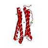 1fhaS S: Starting model for refinement |
|---|---|
| Similar structure data |

-Links
 PDBj
PDBj


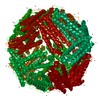
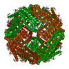
- Assembly
| Deposited unit | 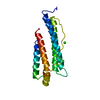
| |||||||||
|---|---|---|---|---|---|---|---|---|---|---|
| 1 | x 24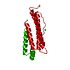
| |||||||||
| Unit cell |
| |||||||||
| Components on special symmetry positions |
|
-Components
| #1: Protein | Mass: 20546.098 Da / Num. of mol.: 1 / Mutation: K82Q, L134P Source method: isolated from a genetically manipulated source Source: (gene. exp.) Rana catesbeiana (American bullfrog) / Strain: BL21-DE3 / Cell line: BL21 / Plasmid: BL21 / Species (production host): Escherichia coli / Production host:  |
|---|---|
| #2: Chemical | ChemComp-CA /   Mass: 40.078 Da / Num. of mol.: 1 / Source method: obtained synthetically / Formula: Ca Mass: 40.078 Da / Num. of mol.: 1 / Source method: obtained synthetically / Formula: Ca |
| #3: Water | ChemComp-HOH /  Mass: 18.015 Da / Num. of mol.: 32 / Source method: isolated from a natural source / Formula: H2O Mass: 18.015 Da / Num. of mol.: 32 / Source method: isolated from a natural source / Formula: H2O |
-Experimental details
-Experiment
| Experiment | Method: X-RAY DIFFRACTION / Number of used crystals: 1 |
|---|
- Sample preparation
| Crystal | Density Matthews: 3.6 Å3/Da / Density % sol: 63 % | ||||||||||||||||||||||||||||||
|---|---|---|---|---|---|---|---|---|---|---|---|---|---|---|---|---|---|---|---|---|---|---|---|---|---|---|---|---|---|---|---|
| Crystal grow | pH: 4.6 / Details: pH 4.6 | ||||||||||||||||||||||||||||||
| Crystal grow | *PLUS Method: vapor diffusion, hanging drop | ||||||||||||||||||||||||||||||
| Components of the solutions | *PLUS
|
-Data collection
| Diffraction | Mean temperature: 120 K |
|---|---|
| Diffraction source | Source: SYNCHROTRON / Site: NSLS  / Beamline: X12C / Wavelength: 1.1 / Beamline: X12C / Wavelength: 1.1 |
| Detector | Type: SIEMENS / Detector: CCD AREA DETECTOR / Date: Feb 1, 1998 / Details: COLLIMATOR |
| Radiation | Monochromator: SI(111) / Monochromatic (M) / Laue (L): M / Scattering type: x-ray |
| Radiation wavelength | Wavelength: 1.1 Å / Relative weight: 1 |
| Reflection | Highest resolution: 1.8 Å / Num. obs: 1179393 / % possible obs: 99.2 % / Observed criterion σ(I): 2 / Redundancy: 60 % / Biso Wilson estimate: 30.5 Å2 / Rmerge(I) obs: 0.078 / Rsym value: 0.078 / Net I/σ(I): 37 |
| Reflection shell | Resolution: 1.81→1.89 Å / Redundancy: 50 % / Rmerge(I) obs: 0.107 / Mean I/σ(I) obs: 0.8 / Rsym value: 0.107 / % possible all: 98.1 |
| Reflection | *PLUS Num. measured all: 1179393 |
| Reflection shell | *PLUS % possible obs: 98.1 % |
- Processing
| Software |
| ||||||||||||||||||||||||||||||||||||||||||||||||||||||||||||||||||||||||||||||||
|---|---|---|---|---|---|---|---|---|---|---|---|---|---|---|---|---|---|---|---|---|---|---|---|---|---|---|---|---|---|---|---|---|---|---|---|---|---|---|---|---|---|---|---|---|---|---|---|---|---|---|---|---|---|---|---|---|---|---|---|---|---|---|---|---|---|---|---|---|---|---|---|---|---|---|---|---|---|---|---|---|---|
| Refinement | Method to determine structure: MOLECULAR REPLACEMENT Starting model: 1FHA Resolution: 1.85→40 Å / Rfactor Rfree error: 0.017 / Data cutoff high absF: 100000 / Data cutoff low absF: 0.001 / Isotropic thermal model: RESTRAINTS / Cross valid method: THROUGHOUT / σ(F): 2
| ||||||||||||||||||||||||||||||||||||||||||||||||||||||||||||||||||||||||||||||||
| Displacement parameters | Biso mean: 30.3 Å2
| ||||||||||||||||||||||||||||||||||||||||||||||||||||||||||||||||||||||||||||||||
| Refine analyze | Luzzati coordinate error obs: 0.02 Å / Luzzati d res low obs: 40 Å | ||||||||||||||||||||||||||||||||||||||||||||||||||||||||||||||||||||||||||||||||
| Refinement step | Cycle: LAST / Resolution: 1.85→40 Å
| ||||||||||||||||||||||||||||||||||||||||||||||||||||||||||||||||||||||||||||||||
| Refine LS restraints |
| ||||||||||||||||||||||||||||||||||||||||||||||||||||||||||||||||||||||||||||||||
| LS refinement shell | Resolution: 1.85→1.93 Å / Rfactor Rfree error: 0.042 / Total num. of bins used: 8
| ||||||||||||||||||||||||||||||||||||||||||||||||||||||||||||||||||||||||||||||||
| Xplor file |
| ||||||||||||||||||||||||||||||||||||||||||||||||||||||||||||||||||||||||||||||||
| Software | *PLUS Name: X-PLOR / Version: 3.8 / Classification: refinement | ||||||||||||||||||||||||||||||||||||||||||||||||||||||||||||||||||||||||||||||||
| Refine LS restraints | *PLUS
|
