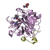
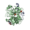
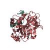
Entry Database : PDB / ID : 3gicTitle Structure of thrombin mutant delta(146-149e) in the free form Thrombin heavy chain Thrombin light chain Keywords / / / / / / / / / / / / / Function / homology Function Domain/homology Component
/ / / / / / / / / / / / / / / / / / / / / / / / / / / / / / / / / / / / / / / / / / / / / / / / / / / / / / / / / / / / / / / / / / / / / / / / / / / / / / / / / / / / / / / / / / / / / / / / / / / / / / / / / / / / / / / / / Biological species Homo sapiens (human)Method / / / / Resolution : 1.55 Å Authors Bah, A. / Carrell, C.J. / Chen, Z. / Gandhi, P.S. / Di Cera, E. Journal : J.Biol.Chem. / Year : 2009Title : Stabilization of the E* form turns thrombin into an anticoagulant.Authors : Bah, A. / Carrell, C.J. / Chen, Z. / Gandhi, P.S. / Di Cera, E. History Deposition Mar 5, 2009 Deposition site / Processing site Revision 1.0 Jun 2, 2009 Provider / Type Revision 1.1 Jul 13, 2011 Group / Version format complianceRevision 1.2 Nov 1, 2017 Group / Category Revision 1.3 Jul 29, 2020 Group Data collection / Database references ... Data collection / Database references / Derived calculations / Structure summary Category chem_comp / entity ... chem_comp / entity / pdbx_chem_comp_identifier / pdbx_entity_nonpoly / struct_ref_seq_dif / struct_site / struct_site_gen Item _chem_comp.name / _chem_comp.type ... _chem_comp.name / _chem_comp.type / _entity.pdbx_description / _pdbx_entity_nonpoly.name / _struct_ref_seq_dif.details Description / Provider / Type Revision 1.4 Sep 6, 2023 Group Data collection / Database references ... Data collection / Database references / Refinement description / Structure summary Category chem_comp / chem_comp_atom ... chem_comp / chem_comp_atom / chem_comp_bond / database_2 / pdbx_initial_refinement_model Item / _database_2.pdbx_DOI / _database_2.pdbx_database_accessionRevision 1.5 Nov 27, 2024 Group / Category / pdbx_modification_feature
Show all Show less
 Movie
Movie Controller


 Open data
 Basic information
 Components
 Keywords
 Function and homology information
 Homo sapiens (human)
 X-RAY DIFFRACTION /
  Authors
 Citation
 Structure visualization
 Downloads & links
 Other downloads
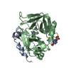


 PDBj
PDBj


 Assembly

 Cricetulus griseus (Chinese hamster) / References: UniProt: P00734, thrombin
 Type: D-saccharide, beta linking / Mass: 221.208 Da / Num. of mol.: 1
 Mass: 18.015 Da / Num. of mol.: 257 / Source method: isolated from a natural source / Formula: H2O
 Sample preparation
 / Beamline: 14-BM-C / Wavelength: 0.9002 Å
 Processing