| 登録情報 | データベース: PDB / ID: 3gic
|
|---|
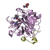
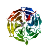
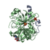
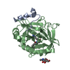
| タイトル | Structure of thrombin mutant delta(146-149e) in the free form |
|---|
 要素 要素 | - Thrombin heavy chain
 トロンビン トロンビン - Thrombin light chainトロンビン
|
|---|
 キーワード キーワード | HYDROLASE (加水分解酵素) / serine protease (セリンプロテアーゼ) / chymotrypsin fold / Acute phase (急性期タンパク質) / Blood coagulation (凝固・線溶系) / Cleavage on pair of basic residues / Disease mutation / Disulfide bond (ジスルフィド) / Gamma-carboxyglutamic acid (Γ-カルボキシグルタミン酸) / Glycoprotein (糖タンパク質) / Kringle / Protease (プロテアーゼ) / Secreted (分泌) / Zymogen (酵素前駆体) |
|---|
| 機能・相同性 |  機能・相同性情報 機能・相同性情報
positive regulation of lipid kinase activity / positive regulation of phospholipase C-activating G protein-coupled receptor signaling pathway / cytolysis by host of symbiont cells / thrombospondin receptor activity / Defective factor XII causes hereditary angioedema / トロンビン / neutrophil-mediated killing of gram-negative bacterium / regulation of blood coagulation / ligand-gated ion channel signaling pathway / Defective F8 cleavage by thrombin ...positive regulation of lipid kinase activity / positive regulation of phospholipase C-activating G protein-coupled receptor signaling pathway / cytolysis by host of symbiont cells / thrombospondin receptor activity / Defective factor XII causes hereditary angioedema / トロンビン / neutrophil-mediated killing of gram-negative bacterium / regulation of blood coagulation / ligand-gated ion channel signaling pathway / Defective F8 cleavage by thrombin / Platelet Aggregation (Plug Formation) / negative regulation of astrocyte differentiation / negative regulation of platelet activation / positive regulation of collagen biosynthetic process / negative regulation of cytokine production involved in inflammatory response / positive regulation of blood coagulation / negative regulation of fibrinolysis / Gamma-carboxylation of protein precursors / Transport of gamma-carboxylated protein precursors from the endoplasmic reticulum to the Golgi apparatus / Common Pathway of Fibrin Clot Formation / Removal of aminoterminal propeptides from gamma-carboxylated proteins / regulation of cytosolic calcium ion concentration / fibrinolysis / Intrinsic Pathway of Fibrin Clot Formation / Peptide ligand-binding receptors / positive regulation of release of sequestered calcium ion into cytosol / Regulation of Complement cascade / acute-phase response / Cell surface interactions at the vascular wall / lipopolysaccharide binding / negative regulation of proteolysis / positive regulation of receptor signaling pathway via JAK-STAT / growth factor activity / positive regulation of insulin secretion / 凝固・線溶系 / response to wounding / Golgi lumen / positive regulation of protein localization to nucleus / Regulation of Insulin-like Growth Factor (IGF) transport and uptake by Insulin-like Growth Factor Binding Proteins (IGFBPs) / positive regulation of reactive oxygen species metabolic process / 凝固・線溶系 / antimicrobial humoral immune response mediated by antimicrobial peptide / Thrombin signalling through proteinase activated receptors (PARs) / heparin binding / regulation of cell shape / positive regulation of cell growth / G alpha (q) signalling events / collagen-containing extracellular matrix / blood microparticle / cell surface receptor signaling pathway / positive regulation of phosphatidylinositol 3-kinase/protein kinase B signal transduction / positive regulation of protein phosphorylation / G protein-coupled receptor signaling pathway / 小胞体 / signaling receptor binding / serine-type endopeptidase activity / calcium ion binding / positive regulation of cell population proliferation / タンパク質分解 / extracellular space / extracellular exosome / extracellular region / 細胞膜類似検索 - 分子機能 Prothrombin/thrombin / Thrombin light chain / Thrombin light chain domain superfamily / Thrombin light chain / Kringle domain / Kringle / Kringle, conserved site / Kringle superfamily / Kringle domain signature. / Kringle domain profile. ...Prothrombin/thrombin / Thrombin light chain / Thrombin light chain domain superfamily / Thrombin light chain / Kringle domain / Kringle / Kringle, conserved site / Kringle superfamily / Kringle domain signature. / Kringle domain profile. / Kringle domain / Vitamin K-dependent carboxylation/gamma-carboxyglutamic (GLA) domain / Gamma-carboxyglutamic acid-rich (GLA) domain / Gamma-carboxyglutamic acid-rich (GLA) domain superfamily / Vitamin K-dependent carboxylation domain. / Gla domain profile. / Domain containing Gla (gamma-carboxyglutamate) residues. / Kringle-like fold / Serine proteases, trypsin family, histidine active site / Serine proteases, trypsin family, serine active site / Peptidase S1A, chymotrypsin family / Serine proteases, trypsin family, histidine active site. / Serine proteases, trypsin domain profile. / Serine proteases, trypsin family, serine active site. / Trypsin-like serine protease / Serine proteases, trypsin domain / トリプシン / Trypsin-like serine proteases / Thrombin, subunit H / Peptidase S1, PA clan, chymotrypsin-like fold / Peptidase S1, PA clan / Βバレル / Mainly Beta類似検索 - ドメイン・相同性 |
|---|
| 生物種 |  Homo sapiens (ヒト) Homo sapiens (ヒト) |
|---|
| 手法 | X線回折 / シンクロトロン / 分子置換 / 解像度: 1.55 Å |
|---|
 データ登録者 データ登録者 | Bah, A. / Carrell, C.J. / Chen, Z. / Gandhi, P.S. / Di Cera, E. |
|---|
 引用 引用 | ジャーナル: J.Biol.Chem. / 年: 2009
タイトル: Stabilization of the E* form turns thrombin into an anticoagulant.
著者: Bah, A. / Carrell, C.J. / Chen, Z. / Gandhi, P.S. / Di Cera, E. |
|---|
| 履歴 | | 登録 | 2009年3月5日 | 登録サイト: RCSB / 処理サイト: RCSB |
|---|
| 改定 1.0 | 2009年6月2日 | Provider: repository / タイプ: Initial release |
|---|
| 改定 1.1 | 2011年7月13日 | Group: Non-polymer description / Version format compliance |
|---|
| 改定 1.2 | 2017年11月1日 | Group: Refinement description / カテゴリ: software |
|---|
| 改定 1.3 | 2020年7月29日 | Group: Data collection / Database references ...Data collection / Database references / Derived calculations / Structure summary
カテゴリ: chem_comp / entity ...chem_comp / entity / pdbx_chem_comp_identifier / pdbx_entity_nonpoly / struct_ref_seq_dif / struct_site / struct_site_gen
Item: _chem_comp.name / _chem_comp.type ..._chem_comp.name / _chem_comp.type / _entity.pdbx_description / _pdbx_entity_nonpoly.name / _struct_ref_seq_dif.details
解説: Carbohydrate remediation / Provider: repository / タイプ: Remediation |
|---|
| 改定 1.4 | 2023年9月6日 | Group: Data collection / Database references ...Data collection / Database references / Refinement description / Structure summary
カテゴリ: chem_comp / chem_comp_atom ...chem_comp / chem_comp_atom / chem_comp_bond / database_2 / pdbx_initial_refinement_model
Item: _chem_comp.pdbx_synonyms / _database_2.pdbx_DOI / _database_2.pdbx_database_accession |
|---|
|
|---|
 ムービー
ムービー コントローラー


 データを開く
 基本情報
基本情報 構造の表示
構造の表示 ダウンロードとリンク
 その他のダウンロード
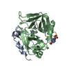


 PDBj
PDBj


 集合体
集合体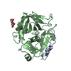


 タイプ: D-saccharide, beta linking / 分子量: 221.208 Da / 分子数: 1 / 由来タイプ: 組換発現 / 式: C8H15NO6
 分子量: 18.015 Da / 分子数: 257 / 由来タイプ: 天然 / 式: H2O
 試料調製
試料調製 / ビームライン: 14-BM-C / 波長: 0.9002 Å
 解析
解析