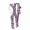
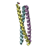
 Movie
Movie Controller


+ Open data

- Basic information
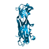
| Entry | Database: PDB / ID: 3g80 | ||||||
|---|---|---|---|---|---|---|---|
| Title | Nodamura virus protein b2, RNA-binding domain | ||||||
 Components Components | Protein B2 | ||||||
 Keywords Keywords | VIRAL PROTEIN / RNA-binding / suppressor of RNAi / RNA interference | ||||||
| Function / homology | ESAT-6-like / Helix Hairpins / RNA binding / Orthogonal Bundle / Mainly Alpha / Protein B2 Function and homology information Function and homology information | ||||||
| Biological species |  Nodamura virus Nodamura virus | ||||||
| Method |  X-RAY DIFFRACTION / SYNCHROTRON / MOLECULAR REPLACEMENT / molecular replacement / Resolution: 2.5 Å X-RAY DIFFRACTION / SYNCHROTRON / MOLECULAR REPLACEMENT / molecular replacement / Resolution: 2.5 Å | ||||||
 Authors Authors | Korber, S. / Shaik Syed Ali, P. / Chen, J.C. | ||||||
 Citation Citation | Journal: Biochemistry / Year: 2009 Title: Structure of the RNA-Binding Domain of Nodamura Virus Protein B2, a Suppressor of RNA Interference. Authors: Korber, S. / Shaik Syed Ali, P. / Chen, J.C. | ||||||
| History |
|
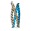
- Structure visualization
| Structure viewer | Molecule: MolmilJmol/JSmol |
|---|
- Downloads & links
-Download
| PDBx/mmCIF format | 3g80.cif.gz | 42.5 KB | Display | PDBx/mmCIF format |
|---|---|---|---|---|
| PDB format | pdb3g80.ent.gz | 29.2 KB | Display | PDB format |
| PDBx/mmJSON format | 3g80.json.gz | Tree view | PDBx/mmJSON format | |
| Others |  Other downloads Other downloads |
-Validation report
| Summary document | 3g80_validation.pdf.gz | 443.7 KB | Display | wwPDB validaton report |
|---|---|---|---|---|
| Full document | 3g80_full_validation.pdf.gz | 447.6 KB | Display | |
| Data in XML | 3g80_validation.xml.gz | 9 KB | Display | |
| Data in CIF | 3g80_validation.cif.gz | 11.7 KB | Display | |
| Arichive directory | https://data.pdbj.org/pub/pdb/validation_reports/g8/3g80ftp://data.pdbj.org/pub/pdb/validation_reports/g8/3g80 | HTTPS FTP |
-Related structure data
| Related structure data | 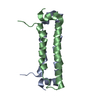 2b9zS S: Starting model for refinement |
|---|---|
| Similar structure data |


-Links
 PDBj
PDBj- Assembly
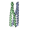
| Deposited unit | 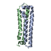
| ||||||||
|---|---|---|---|---|---|---|---|---|---|
| 1 |
| ||||||||
| Unit cell |
|
-Components
| #1: Protein | Mass: 11136.767 Da / Num. of mol.: 2 / Fragment: RNA-binding domain Source method: isolated from a genetically manipulated source Source: (gene. exp.) Nodamura virus / Gene: B2 / Production host:  #2: Water | ChemComp-HOH / |  Mass: 18.015 Da / Num. of mol.: 81 / Source method: isolated from a natural source / Formula: H2O Mass: 18.015 Da / Num. of mol.: 81 / Source method: isolated from a natural source / Formula: H2O |
|---|
-Experimental details
-Experiment
| Experiment | Method: X-RAY DIFFRACTION / Number of used crystals: 1 |
|---|
- Sample preparation
| Crystal | Density Matthews: 2.02 Å3/Da / Density % sol: 38.97 % |
|---|---|
| Crystal grow | Temperature: 298 K / Method: vapor diffusion, sitting drop / pH: 8.5 Details: 15% PEG 3350, pH 8.5, VAPOR DIFFUSION, SITTING DROP, temperature 298K |
-Data collection
| Diffraction | Mean temperature: 100 K |
|---|---|
| Diffraction source | Source: SYNCHROTRON / Site: ESRF  / Beamline: ID29 / Wavelength: 0.9762 Å / Beamline: ID29 / Wavelength: 0.9762 Å |
| Detector | Type: ADSC QUANTUM 210 / Detector: CCD / Date: Sep 3, 2008 |
| Radiation | Monochromator: Si(311) / Protocol: SINGLE WAVELENGTH / Monochromatic (M) / Laue (L): M / Scattering type: x-ray |
| Radiation wavelength | Wavelength: 0.9762 Å / Relative weight: 1 |
| Reflection | Resolution: 2.5→500 Å / Num. all: 6658 / Num. obs: 6576 / % possible obs: 99.2 % / Observed criterion σ(F): 0 / Observed criterion σ(I): 0 / Redundancy: 6.3 % / Biso Wilson estimate: 34 Å2 / Rmerge(I) obs: 0.097 / Rsym value: 0.097 / Net I/σ(I): 6.3 |
| Reflection shell | Resolution: 2.5→2.64 Å / Rmerge(I) obs: 0.428 / Mean I/σ(I) obs: 1.7 / Rsym value: 0.428 / % possible all: 100 |
-Phasing
| Phasing | Method: molecular replacement |
|---|
- Processing
| Software |
| ||||||||||||||||||||||||||||
|---|---|---|---|---|---|---|---|---|---|---|---|---|---|---|---|---|---|---|---|---|---|---|---|---|---|---|---|---|---|
| Refinement | Method to determine structure: MOLECULAR REPLACEMENT Starting model: 2B9Z.pdb Resolution: 2.5→500 Å / Occupancy max: 1 / Occupancy min: 1 / Cross valid method: THROUGHOUT / σ(F): 0 / σ(I): 0 / Stereochemistry target values: Engh & Huber
| ||||||||||||||||||||||||||||
| Solvent computation | Bsol: 69.78 Å2 | ||||||||||||||||||||||||||||
| Displacement parameters | Biso max: 67.87 Å2 / Biso mean: 34.016 Å2 / Biso min: 11.7 Å2
| ||||||||||||||||||||||||||||
| Refine analyze |
| ||||||||||||||||||||||||||||
| Refinement step | Cycle: LAST / Resolution: 2.5→500 Å
| ||||||||||||||||||||||||||||
| Refine LS restraints |
| ||||||||||||||||||||||||||||
| Xplor file |
|
