 Movie
Movie Controller

[English] 日本語
 Yorodumi
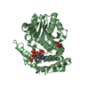
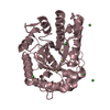
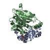
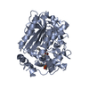
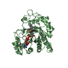
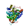
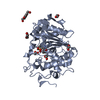
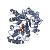
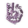
- PDB-3g16: Crystal structure of protein of unknown function with cystatin-li... -
+ Open data

- Basic information
| Entry | Database: PDB / ID: 3g16 | ||||||
|---|---|---|---|---|---|---|---|
| Title | Crystal structure of protein of unknown function with cystatin-like fold (YP_001022489.1) from METHYLOBIUM PETROLEOPHILUM PM1 at 1.45 A resolution | ||||||
 Components Components | uncharacterized protein with cystatin-like fold | ||||||
 Keywords Keywords | structural genomics / unknown function / YP_001022489.1 / protein of unknown function with cystatin-like fold / Joint Center for Structural Genomics / JCSG / Protein Structure Initiative / PSI-2 | ||||||
| Function / homology | SnoaL-like domain / SnoaL-like domain / Nuclear Transport Factor 2; Chain: A, - #50 / NTF2-like domain superfamily / Nuclear Transport Factor 2; Chain: A, / Roll / Alpha Beta / Unknown ligand / SnoaL-like domain-containing protein Function and homology information Function and homology information | ||||||
| Biological species |  Methylibium petroleiphilum PM1 (bacteria) Methylibium petroleiphilum PM1 (bacteria) | ||||||
| Method |  X-RAY DIFFRACTION / SYNCHROTRON / MAD / Resolution: 1.45 Å X-RAY DIFFRACTION / SYNCHROTRON / MAD / Resolution: 1.45 Å | ||||||
 Authors Authors | Joint Center for Structural Genomics (JCSG) | ||||||
 Citation Citation | Journal: To be published Title: Crystal structure of protein of unknown function with cystatin-like fold (YP_001022489.1) from METHYLOBIUM PETROLEOPHILUM PM1 at 1.45 A resolution Authors: Joint Center for Structural Genomics (JCSG) | ||||||
| History |
|
- Structure visualization
| Structure viewer | Molecule: MolmilJmol/JSmol |
|---|
- Downloads & links
-Download
| PDBx/mmCIF format | 3g16.cif.gz | 86.2 KB | Display | PDBx/mmCIF format |
|---|---|---|---|---|
| PDB format | pdb3g16.ent.gz | 65.1 KB | Display | PDB format |
| PDBx/mmJSON format | 3g16.json.gz | Tree view | PDBx/mmJSON format | |
| Others |  Other downloads Other downloads |
-Validation report
| Arichive directory | https://data.pdbj.org/pub/pdb/validation_reports/g1/3g16ftp://data.pdbj.org/pub/pdb/validation_reports/g1/3g16 | HTTPS FTP |
|---|
-Related structure data
| Similar structure data | |
|---|---|
| Other databases |

-Links
 PDBj
PDBj


- Assembly
| Deposited unit | 
| ||||||||
|---|---|---|---|---|---|---|---|---|---|
| 1 |
| ||||||||
| Unit cell |
| ||||||||
| Details | CRYSTAL PACKING ANALYSIS AND ANALYTICAL SIZE EXCLUSION CHROMATOGRAPHY SUGGEST THAT A DIMER IS THE STABLE OLIGOMERIC FORM IN SOLUTION. |
-Components
| #1: Protein | Mass: 17472.066 Da / Num. of mol.: 2 Source method: isolated from a genetically manipulated source Source: (gene. exp.) Methylibium petroleiphilum PM1 (bacteria)Gene: Mpe_A3301, YP_001022489.1 / Plasmid: SpeedET / Production host: #2: Chemical | Num. of mol.: 2 / Source method: obtained synthetically #3: Chemical | ChemComp-CL /   Mass: 35.453 Da / Num. of mol.: 4 / Source method: obtained synthetically / Formula: Cl Mass: 35.453 Da / Num. of mol.: 4 / Source method: obtained synthetically / Formula: Cl#4: Chemical |   Mass: 92.094 Da / Num. of mol.: 2 / Source method: obtained synthetically / Formula: C3H8O3 Mass: 92.094 Da / Num. of mol.: 2 / Source method: obtained synthetically / Formula: C3H8O3#5: Water | ChemComp-HOH / |  Mass: 18.015 Da / Num. of mol.: 372 / Source method: isolated from a natural source / Formula: H2O Mass: 18.015 Da / Num. of mol.: 372 / Source method: isolated from a natural source / Formula: H2OHas protein modification | Y | Sequence details | THIS CONSTRUCT WAS EXPRESSED WITH A PURIFICATION TAG MGSDKIHHHHHHENLYFQG. THE TAG WAS REMOVED WITH ...THIS CONSTRUCT WAS EXPRESSED WITH A PURIFICATI | |
|---|
-Experimental details
-Experiment
| Experiment | Method: X-RAY DIFFRACTION / Number of used crystals: 1 |
|---|
- Sample preparation
| Crystal | Density Matthews: 2.56 Å3/Da / Density % sol: 51.87 % |
|---|---|
| Crystal grow | Temperature: 277 K / Method: vapor diffusion, sitting drop / pH: 6 Details: 1.0000M LiCl, 20.0000% PEG-6000, 0.1M MES pH 6.0, NANODROP, VAPOR DIFFUSION, SITTING DROP, temperature 277K |
-Data collection
| Diffraction | Mean temperature: 100 K | |||||||||||||||||||||||||||||||||||||||||||||||||||||||||||||||||||||||||||||
|---|---|---|---|---|---|---|---|---|---|---|---|---|---|---|---|---|---|---|---|---|---|---|---|---|---|---|---|---|---|---|---|---|---|---|---|---|---|---|---|---|---|---|---|---|---|---|---|---|---|---|---|---|---|---|---|---|---|---|---|---|---|---|---|---|---|---|---|---|---|---|---|---|---|---|---|---|---|---|
| Diffraction source | Source: SYNCHROTRON / Site: SSRL  / Beamline: BL11-1 / Wavelength: 0.91837,0.97864,0.97817 / Beamline: BL11-1 / Wavelength: 0.91837,0.97864,0.97817 | |||||||||||||||||||||||||||||||||||||||||||||||||||||||||||||||||||||||||||||
| Detector | Type: MARMOSAIC 325 mm CCD / Detector: CCD / Date: Nov 12, 2008 / Details: Flat mirror (vertical focusing) | |||||||||||||||||||||||||||||||||||||||||||||||||||||||||||||||||||||||||||||
| Radiation | Monochromator: Single crystal Si(111) bent monochromator (horizontal focusing) Protocol: MAD / Monochromatic (M) / Laue (L): M / Scattering type: x-ray | |||||||||||||||||||||||||||||||||||||||||||||||||||||||||||||||||||||||||||||
| Radiation wavelength |
| |||||||||||||||||||||||||||||||||||||||||||||||||||||||||||||||||||||||||||||
| Reflection | Resolution: 1.45→27.973 Å / Num. obs: 63677 / % possible obs: 97.7 % / Observed criterion σ(I): -3 / Biso Wilson estimate: 18.269 Å2 / Rmerge(I) obs: 0.025 / Net I/σ(I): 16.47 | |||||||||||||||||||||||||||||||||||||||||||||||||||||||||||||||||||||||||||||
| Reflection shell |
|
-Phasing
| Phasing | Method: MAD |
|---|
- Processing
| Software |
| |||||||||||||||||||||||||||||||||||||||||||||||||||||||||||||||||||||||||||||||||||||||||||||||||||||||||||||||||||||||||||||
|---|---|---|---|---|---|---|---|---|---|---|---|---|---|---|---|---|---|---|---|---|---|---|---|---|---|---|---|---|---|---|---|---|---|---|---|---|---|---|---|---|---|---|---|---|---|---|---|---|---|---|---|---|---|---|---|---|---|---|---|---|---|---|---|---|---|---|---|---|---|---|---|---|---|---|---|---|---|---|---|---|---|---|---|---|---|---|---|---|---|---|---|---|---|---|---|---|---|---|---|---|---|---|---|---|---|---|---|---|---|---|---|---|---|---|---|---|---|---|---|---|---|---|---|---|---|---|
| Refinement | Method to determine structure: MAD / Resolution: 1.45→27.973 Å / Cor.coef. Fo:Fc: 0.974 / Cor.coef. Fo:Fc free: 0.966 / Occupancy max: 1 / Occupancy min: 0.25 / SU B: 1.695 / SU ML: 0.033 / TLS residual ADP flag: LIKELY RESIDUAL / Cross valid method: THROUGHOUT / σ(F): 0 / ESU R: 0.054 / ESU R Free: 0.054 Stereochemistry target values: MAXIMUM LIKELIHOOD WITH PHASES Details: 1.HYDROGENS HAVE BEEN ADDED IN THE RIDING POSITIONS. 2.ATOM RECORDS CONTAIN RESIDUAL B FACTORS ONLY. 3.A MET-INHIBITION PROTOCOL WAS USED FOR SELENOMETHIONINE INCORPORATION DURING PROTEIN ...Details: 1.HYDROGENS HAVE BEEN ADDED IN THE RIDING POSITIONS. 2.ATOM RECORDS CONTAIN RESIDUAL B FACTORS ONLY. 3.A MET-INHIBITION PROTOCOL WAS USED FOR SELENOMETHIONINE INCORPORATION DURING PROTEIN EXPRESSION. THE OCCUPANCY OF THE SE ATOMS IN THE MSE RESIDUES WAS REDUCED TO 0.75 FOR THE REDUCED SCATTERING POWER DUE TO PARTIAL S-MET INCORPORATION. 4.CHLORIDE (CL) FROM THE CRYSTALLIZATION AND GLYCEROL (GOL) FROM THE CRYOPROTECTION CONDITION HAVE BEEN MODELED IN THE SOLVENT STRUCTURE. 5.AN UNIDENTIFIED LIGAND (UNL) HAS BEEN MODELED IN THE PUTATIVE ACTIVE SITE IN BOTH PROTEIN MOLECULES. IT IS POSSIBLE THAT THIS UNL IS ACTUALLY A PARTIALLY OCCUPIED MES MOLECULE FROM THE CRYSTALLIZATION BUFFER. THIS INFERENCE IS BASED ON THE LOWER B-FACTOR OF ATOM O7 OF UNL-1 WHICH SUGGESTS THAT THIS MAY BE A HEAVIER ATOM (LIKE SULFUR). 6.RESIDUES 45-48 IN CHAIN B HAVE POOR ELECTRON DENSITY AND AND HAVE BEEN MODELED BASED ON THE CORRESPONDING REGION OF CHAIN A.
| |||||||||||||||||||||||||||||||||||||||||||||||||||||||||||||||||||||||||||||||||||||||||||||||||||||||||||||||||||||||||||||
| Solvent computation | Ion probe radii: 0.8 Å / Shrinkage radii: 0.8 Å / VDW probe radii: 1.2 Å / Solvent model: MASK | |||||||||||||||||||||||||||||||||||||||||||||||||||||||||||||||||||||||||||||||||||||||||||||||||||||||||||||||||||||||||||||
| Displacement parameters | Biso max: 68.37 Å2 / Biso mean: 20.701 Å2 / Biso min: 9.07 Å2
| |||||||||||||||||||||||||||||||||||||||||||||||||||||||||||||||||||||||||||||||||||||||||||||||||||||||||||||||||||||||||||||
| Refinement step | Cycle: LAST / Resolution: 1.45→27.973 Å
| |||||||||||||||||||||||||||||||||||||||||||||||||||||||||||||||||||||||||||||||||||||||||||||||||||||||||||||||||||||||||||||
| Refine LS restraints |
| |||||||||||||||||||||||||||||||||||||||||||||||||||||||||||||||||||||||||||||||||||||||||||||||||||||||||||||||||||||||||||||
| LS refinement shell | Resolution: 1.452→1.49 Å / Total num. of bins used: 20
| |||||||||||||||||||||||||||||||||||||||||||||||||||||||||||||||||||||||||||||||||||||||||||||||||||||||||||||||||||||||||||||
| Refinement TLS params. | Method: refined / Refine-ID: X-RAY DIFFRACTION
| |||||||||||||||||||||||||||||||||||||||||||||||||||||||||||||||||||||||||||||||||||||||||||||||||||||||||||||||||||||||||||||
| Refinement TLS group |
|
