 Movie
Movie Controller


+ Open data

- Basic information
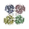
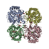
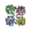
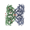
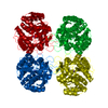
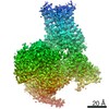
| Entry | Database: PDB / ID: 3fz0 | ||||||
|---|---|---|---|---|---|---|---|
| Title | Inosine-Guanosine Nucleoside Hydrolase (IG-NH) | ||||||
 Components Components | Nucleoside hydrolase, putative | ||||||
 Keywords Keywords | HYDROLASE / NH fold / open alpha/beta structure / Glycosidase | ||||||
| Function / homology |  Function and homology information Function and homology informationguanosine salvage / inosine salvage / purine nucleosidase / inosine nucleosidase activity / glycosome / axoneme / metal ion binding / cytoplasm Similarity search - Function | ||||||
| Biological species |  | ||||||
| Method |  X-RAY DIFFRACTION / SYNCHROTRON / MOLECULAR REPLACEMENT / Resolution: 2.5 Å X-RAY DIFFRACTION / SYNCHROTRON / MOLECULAR REPLACEMENT / Resolution: 2.5 Å | ||||||
 Authors Authors | Vandemeulebroucke, A. / Minici, C. / Bruno, I. / Muzzolini, L. / Tornaghi, P. / Parkin, D.W. / Schramm, V.L. / Versees, W. / Steyaert, J. / Degano, M. | ||||||
 Citation Citation | Journal: Biochemistry / Year: 2010 Title: Structure and mechanism of the 6-oxopurine nucleosidase from Trypanosoma brucei brucei Authors: Vandemeulebroucke, A. / Minici, C. / Bruno, I. / Muzzolini, L. / Tornaghi, P. / Parkin, D.W. / Versees, W. / Steyaert, J. / Degano, M. #1: Journal: J.Biol.Chem. / Year: 1994 Title: Guanosine-inosine-preferring nucleoside N-glycohydrolase from Crithidia fasciculata Authors: Estupinan, B. / Schramm, V.L. | ||||||
| History |
|
- Structure visualization
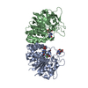
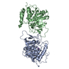
| Structure viewer | Molecule: MolmilJmol/JSmol |
|---|
- Downloads & links
-Download
| PDBx/mmCIF format | 3fz0.cif.gz | 267.8 KB | Display | PDBx/mmCIF format |
|---|---|---|---|---|
| PDB format | pdb3fz0.ent.gz | 216.4 KB | Display | PDB format |
| PDBx/mmJSON format | 3fz0.json.gz | Tree view | PDBx/mmJSON format | |
| Others |  Other downloads Other downloads |
-Validation report
| Arichive directory | https://data.pdbj.org/pub/pdb/validation_reports/fz/3fz0ftp://data.pdbj.org/pub/pdb/validation_reports/fz/3fz0 | HTTPS FTP |
|---|
-Related structure data
| Related structure data | 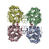 1q8fS S: Starting model for refinement |
|---|---|
| Similar structure data |
-Links
 PDBj
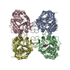
PDBj- Assembly
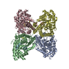
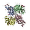
| Deposited unit | 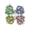
| ||||||||||||||||||||||||||||||||||||||||||||||||||||||||||||||||||||||||||||||||||||||||||||||||||||||||||||||||||||||||||||||||||||||||||||||||||||||||||||||||||||||||||||||||
|---|---|---|---|---|---|---|---|---|---|---|---|---|---|---|---|---|---|---|---|---|---|---|---|---|---|---|---|---|---|---|---|---|---|---|---|---|---|---|---|---|---|---|---|---|---|---|---|---|---|---|---|---|---|---|---|---|---|---|---|---|---|---|---|---|---|---|---|---|---|---|---|---|---|---|---|---|---|---|---|---|---|---|---|---|---|---|---|---|---|---|---|---|---|---|---|---|---|---|---|---|---|---|---|---|---|---|---|---|---|---|---|---|---|---|---|---|---|---|---|---|---|---|---|---|---|---|---|---|---|---|---|---|---|---|---|---|---|---|---|---|---|---|---|---|---|---|---|---|---|---|---|---|---|---|---|---|---|---|---|---|---|---|---|---|---|---|---|---|---|---|---|---|---|---|---|---|---|
| 1 |
| ||||||||||||||||||||||||||||||||||||||||||||||||||||||||||||||||||||||||||||||||||||||||||||||||||||||||||||||||||||||||||||||||||||||||||||||||||||||||||||||||||||||||||||||||
| Unit cell |
| ||||||||||||||||||||||||||||||||||||||||||||||||||||||||||||||||||||||||||||||||||||||||||||||||||||||||||||||||||||||||||||||||||||||||||||||||||||||||||||||||||||||||||||||||
| Noncrystallographic symmetry (NCS) | NCS domain:
NCS domain segments: Refine code: 1
|