 Movie
Movie Controller

[English] 日本語
 Yorodumi
- PDB-1q8f: Crystal Structure of the E.coli pyrimidine nucleoside hydrolase yeiK -
+ Open data

- Basic information
| Entry | Database: PDB / ID: 1q8f | ||||||
|---|---|---|---|---|---|---|---|
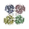
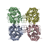
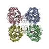
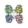
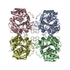
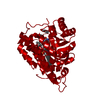
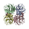
| Title | Crystal Structure of the E.coli pyrimidine nucleoside hydrolase yeiK | ||||||
 Components Components | Pyrimidine nucleoside hydrolase | ||||||
 Keywords Keywords | HYDROLASE / open alpha-beta structure / NH-fold | ||||||
| Function / homology |  Function and homology information Function and homology informationribosylpyrimidine nucleosidase / pyrimidine ribonucleoside catabolic process / uridine nucleosidase activity / purine nucleosidase activity / pyrimidine nucleobase metabolic process / purine nucleoside catabolic process / protein homotetramerization / calcium ion binding / protein-containing complex / identical protein binding / cytosol Similarity search - Function | ||||||
| Biological species |  | ||||||
| Method |  X-RAY DIFFRACTION / SYNCHROTRON / MOLECULAR REPLACEMENT / Resolution: 1.7 Å X-RAY DIFFRACTION / SYNCHROTRON / MOLECULAR REPLACEMENT / Resolution: 1.7 Å | ||||||
 Authors Authors | Giabbai, B. / Degano, M. | ||||||
 Citation Citation | Journal: STRUCTURE / Year: 2004 Title: Crystal structure to 1.7 a of the Escherichia coli pyrimidine nucleoside hydrolase YeiK, a novel candidate for cancer gene therapy. Authors: Giabbai, B. / Degano, M. #1: Journal: Biochemistry / Year: 1996Title: Three-dimensional structure of the Inosine-Uridine Nucleoside N-ribohydrolase from Crithidia fasciculata Authors: Degano, M. / Gopaul, D.N. / Scapin, G. / Schramm, V.L. / Sacchettini, J.C. #2: Journal: Biochemistry / Year: 1998Title: Trypanosomal nucleoside hydrolase. A novel mechanism from the structure with a transition-state inhibitor Authors: Degano, M. / Almo, S.C. / Sacchettini, J.C. / Schramm, V.L. #3: Journal: J.Mol.Biol. / Year: 2001Title: Structure and function of a novel purine specific nucleoside hydrolase from Trypanosoma vivax Authors: Versees, W. / Decanniere, K. / Pelle, R. / Depoorter, J. / Brosens, E. / Parkin, D.W. / Steyaert, J. | ||||||
| History |
|
- Structure visualization
| Structure viewer | Molecule: MolmilJmol/JSmol |
|---|
- Downloads & links
-Download
| PDBx/mmCIF format | 1q8f.cif.gz | 266.1 KB | Display | PDBx/mmCIF format |
|---|---|---|---|---|
| PDB format | pdb1q8f.ent.gz | 211 KB | Display | PDB format |
| PDBx/mmJSON format | 1q8f.json.gz | Tree view | PDBx/mmJSON format | |
| Others |  Other downloads Other downloads |
-Validation report
| Arichive directory | https://data.pdbj.org/pub/pdb/validation_reports/q8/1q8fftp://data.pdbj.org/pub/pdb/validation_reports/q8/1q8f | HTTPS FTP |
|---|
-Related structure data
| Related structure data | 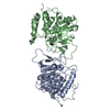 1masS S: Starting model for refinement |
|---|---|
| Similar structure data |


-Links
 PDBj
PDBj- Assembly
| Deposited unit | 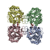
| |||||||||||||||||||||||||||||||||||||||||||||||||||||||||||||||||||||||||||||||||||||||||||||||||||||||||||||||||||||||||||||||||||||||||||||||||||||||||||||||||||||||||||||||||||||||||||||||||||||||||||||||||||||||||||||||||||||
|---|---|---|---|---|---|---|---|---|---|---|---|---|---|---|---|---|---|---|---|---|---|---|---|---|---|---|---|---|---|---|---|---|---|---|---|---|---|---|---|---|---|---|---|---|---|---|---|---|---|---|---|---|---|---|---|---|---|---|---|---|---|---|---|---|---|---|---|---|---|---|---|---|---|---|---|---|---|---|---|---|---|---|---|---|---|---|---|---|---|---|---|---|---|---|---|---|---|---|---|---|---|---|---|---|---|---|---|---|---|---|---|---|---|---|---|---|---|---|---|---|---|---|---|---|---|---|---|---|---|---|---|---|---|---|---|---|---|---|---|---|---|---|---|---|---|---|---|---|---|---|---|---|---|---|---|---|---|---|---|---|---|---|---|---|---|---|---|---|---|---|---|---|---|---|---|---|---|---|---|---|---|---|---|---|---|---|---|---|---|---|---|---|---|---|---|---|---|---|---|---|---|---|---|---|---|---|---|---|---|---|---|---|---|---|---|---|---|---|---|---|---|---|---|---|---|---|---|---|---|---|
| 1 |
| |||||||||||||||||||||||||||||||||||||||||||||||||||||||||||||||||||||||||||||||||||||||||||||||||||||||||||||||||||||||||||||||||||||||||||||||||||||||||||||||||||||||||||||||||||||||||||||||||||||||||||||||||||||||||||||||||||||
| Unit cell |
| |||||||||||||||||||||||||||||||||||||||||||||||||||||||||||||||||||||||||||||||||||||||||||||||||||||||||||||||||||||||||||||||||||||||||||||||||||||||||||||||||||||||||||||||||||||||||||||||||||||||||||||||||||||||||||||||||||||
| Noncrystallographic symmetry (NCS) | NCS domain:
NCS domain segments:
|