 Movie
Movie Controller


+ Open data

- Basic information
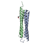
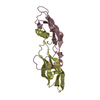
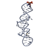
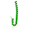
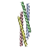
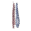
| Entry | Database: PDB / ID: 3frv | ||||||
|---|---|---|---|---|---|---|---|
| Title | Structure of Human CHMP3 (residues 1-150) | ||||||
 Components Components | Charged multivesicular body protein 3 | ||||||
 Keywords Keywords | PROTEIN TRANSPORT / ESCRT / ESCRT-III / CHMP / IST1 / Coiled coil / Cytoplasm / Lipoprotein / Membrane / Myristate / Phosphoprotein / Transport | ||||||
| Function / homology |  Function and homology information Function and homology informationregulation of endosome size / multivesicular body-lysosome fusion / amphisome membrane / vesicle fusion with vacuole / late endosome to lysosome transport / ESCRT III complex / kinetochore microtubule / endosome transport via multivesicular body sorting pathway / nuclear membrane reassembly / multivesicular body sorting pathway ...regulation of endosome size / multivesicular body-lysosome fusion / amphisome membrane / vesicle fusion with vacuole / late endosome to lysosome transport / ESCRT III complex / kinetochore microtubule / endosome transport via multivesicular body sorting pathway / nuclear membrane reassembly / multivesicular body sorting pathway / Sealing of the nuclear envelope (NE) by ESCRT-III / suppression of viral release by host / midbody abscission / regulation of centrosome duplication / membrane fission / late endosome to vacuole transport / multivesicular body assembly / multivesicular body membrane / phosphatidylcholine binding / ubiquitin-dependent protein catabolic process via the multivesicular body sorting pathway / plasma membrane repair / regulation of mitotic spindle assembly / Translation of Replicase and Assembly of the Replication Transcription Complex / nucleus organization / molecular function inhibitor activity / regulation of early endosome to late endosome transport / Macroautophagy / positive regulation of cytokinesis / viral budding via host ESCRT complex / ubiquitin-specific protease binding / mitotic metaphase chromosome alignment / autophagosome membrane / viral release from host cell / protein polymerization / Pyroptosis / nuclear pore / autophagosome maturation / multivesicular body / phosphatidylinositol-4,5-bisphosphate binding / Endosomal Sorting Complex Required For Transport (ESCRT) / viral budding from plasma membrane / HCMV Late Events / macroautophagy / Late endosomal microautophagy / Budding and maturation of HIV virion / kinetochore / autophagy / late endosome / protein transport / cytoplasmic vesicle / Translation of Replicase and Assembly of the Replication Transcription Complex / midbody / early endosome / lysosomal membrane / apoptotic process / extracellular exosome / identical protein binding / plasma membrane / cytosol Similarity search - Function | ||||||
| Biological species |  Homo sapiens (human) Homo sapiens (human) | ||||||
| Method |  X-RAY DIFFRACTION / MOLECULAR REPLACEMENT / Resolution: 3.7 Å X-RAY DIFFRACTION / MOLECULAR REPLACEMENT / Resolution: 3.7 Å | ||||||
 Authors Authors | Hill, C.P. / Schubert, H.L. / McCullough, J. / Sundquist, W.I. | ||||||
 Citation Citation | Journal: Nat.Struct.Mol.Biol. / Year: 2009 Title: Structural basis for ESCRT-III protein autoinhibition. Authors: Bajorek, M. / Schubert, H.L. / McCullough, J. / Langelier, C. / Eckert, D.M. / Stubblefield, W.M. / Uter, N.T. / Myszka, D.G. / Hill, C.P. / Sundquist, W.I. | ||||||
| History |
|
- Structure visualization
| Structure viewer | Molecule: MolmilJmol/JSmol |
|---|
- Downloads & links
-Download
| PDBx/mmCIF format | 3frv.cif.gz | 37.7 KB | Display | PDBx/mmCIF format |
|---|---|---|---|---|
| PDB format | pdb3frv.ent.gz | 25.9 KB | Display | PDB format |
| PDBx/mmJSON format | 3frv.json.gz | Tree view | PDBx/mmJSON format | |
| Others |  Other downloads Other downloads |
-Validation report
| Arichive directory | https://data.pdbj.org/pub/pdb/validation_reports/fr/3frvftp://data.pdbj.org/pub/pdb/validation_reports/fr/3frv | HTTPS FTP |
|---|
-Related structure data
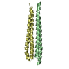
| Related structure data |  3frrC  3frsC 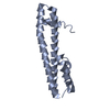 3frtC 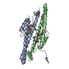 2gd5S C: citing same article ( S: Starting model for refinement |
|---|---|
| Similar structure data |


-Links
 PDBj
PDBj


- Assembly
| Deposited unit | 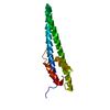
| ||||||||
|---|---|---|---|---|---|---|---|---|---|
| 1 |
| ||||||||
| Unit cell |
|
-Components
| #1: Protein | Mass: 17408.699 Da / Num. of mol.: 1 / Fragment: UNP residues 1 - 150 Source method: isolated from a genetically manipulated source Source: (gene. exp.) Homo sapiens (human) / Gene: CGI-149, CHMP3, NEDF, VPS24 / Plasmid: pGST / Production host:  |
|---|
-Experimental details
-Experiment
| Experiment | Method: X-RAY DIFFRACTION / Number of used crystals: 1 |
|---|
- Sample preparation
| Crystal | Density Matthews: 3.15 Å3/Da / Density % sol: 60.95 % |
|---|---|
| Crystal grow | Temperature: 286 K / Method: vapor diffusion, sitting drop / pH: 7.5 Details: 16% PEG 3350, 100 mM HEPES, 200mM Proline, pH 7.5, VAPOR DIFFUSION, SITTING DROP, temperature 286K |
-Data collection
| Diffraction | Mean temperature: 100 K |
|---|---|
| Diffraction source | Source: ROTATING ANODE / Type: RIGAKU MICROMAX-007 HF / Wavelength: 1.54 Å |
| Detector | Type: RIGAKU RAXIS IV / Detector: IMAGE PLATE / Date: Nov 25, 2008 / Details: Verimax HR |
| Radiation | Monochromator: verimax HR / Protocol: SINGLE WAVELENGTH / Monochromatic (M) / Laue (L): M / Scattering type: x-ray |
| Radiation wavelength | Wavelength: 1.54 Å / Relative weight: 1 |
| Reflection | Resolution: 3.7→25 Å / Num. all: 2586 / Num. obs: 2451 / % possible obs: 99.2 % / Observed criterion σ(F): 0 / Observed criterion σ(I): 0 / Redundancy: 3.2 % / Biso Wilson estimate: 101 Å2 / Rmerge(I) obs: 0.63 / Net I/σ(I): 18.3 |
| Reflection shell | Resolution: 3.7→3.83 Å / Redundancy: 3.1 % / Rmerge(I) obs: 0.553 / Mean I/σ(I) obs: 2.2 / % possible all: 99.2 |
- Processing
| Software |
| ||||||||||||||||||||
|---|---|---|---|---|---|---|---|---|---|---|---|---|---|---|---|---|---|---|---|---|---|
| Refinement | Method to determine structure: MOLECULAR REPLACEMENT Starting model: PDB ENTRY 2GD5 Resolution: 3.7→25 Å / σ(F): 0 / Stereochemistry target values: Engh & Huber Details: rigid body only - against anisotropically adjusted Fs by PHASER.
| ||||||||||||||||||||
| Refinement step | Cycle: LAST / Resolution: 3.7→25 Å
|
