 Movie
Movie Controller


+ Open data

- Basic information
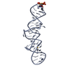
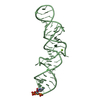
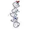
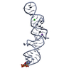
| Entry | Database: PDB / ID: 2gyr | ||||||
|---|---|---|---|---|---|---|---|
| Title | Crystal structure of human artemin | ||||||
 Components Components | Neurotrophic factor artemin, isoform 3 | ||||||
 Keywords Keywords | HORMONE/GROWTH FACTOR / neurotrophic factor / cystein-knot / HORMONE-GROWTH FACTOR COMPLEX | ||||||
| Function / homology |  Function and homology information Function and homology informationglial cell-derived neurotrophic factor receptor binding / Peyer's patch morphogenesis / glial cell-derived neurotrophic factor receptor signaling pathway / lymphocyte migration into lymphoid organs / induction of positive chemotaxis / peripheral nervous system development / NCAM1 interactions / neuroblast proliferation / RET signaling / axon guidance ...glial cell-derived neurotrophic factor receptor binding / Peyer's patch morphogenesis / glial cell-derived neurotrophic factor receptor signaling pathway / lymphocyte migration into lymphoid organs / induction of positive chemotaxis / peripheral nervous system development / NCAM1 interactions / neuroblast proliferation / RET signaling / axon guidance / growth factor activity / receptor tyrosine kinase binding / RAF/MAP kinase cascade / signaling receptor binding / signal transduction / : / extracellular region Similarity search - Function | ||||||
| Biological species |  Homo sapiens (human) Homo sapiens (human) | ||||||
| Method |  X-RAY DIFFRACTION / SYNCHROTRON / SIRAS / Resolution: 2.6 Å X-RAY DIFFRACTION / SYNCHROTRON / SIRAS / Resolution: 2.6 Å | ||||||
 Authors Authors | Wang, X.Q. | ||||||
 Citation Citation | Journal: Structure / Year: 2006 Title: Structure of Artemin Complexed with Its Receptor GFRalpha3: Convergent Recognition of Glial Cell Line-Derived Neurotrophic Factors. Authors: Wang, X. / Baloh, R.H. / Milbrandt, J. / Garcia, K.C. | ||||||
| History |
|
- Structure visualization
| Structure viewer | Molecule: MolmilJmol/JSmol |
|---|
- Downloads & links
-Download
| PDBx/mmCIF format | 2gyr.cif.gz | 115 KB | Display | PDBx/mmCIF format |
|---|---|---|---|---|
| PDB format | pdb2gyr.ent.gz | 92.8 KB | Display | PDB format |
| PDBx/mmJSON format | 2gyr.json.gz | Tree view | PDBx/mmJSON format | |
| Others |  Other downloads Other downloads |
-Validation report
| Arichive directory | https://data.pdbj.org/pub/pdb/validation_reports/gy/2gyrftp://data.pdbj.org/pub/pdb/validation_reports/gy/2gyr | HTTPS FTP |
|---|
-Related structure data


-Links
 PDBj
PDBj


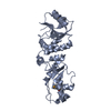
- Assembly
| Deposited unit | 
| ||||||||
|---|---|---|---|---|---|---|---|---|---|
| 1 | 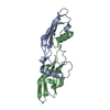
| ||||||||
| 2 | 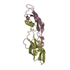
| ||||||||
| 3 | 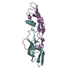
| ||||||||
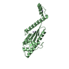
| Unit cell |
|
-Components
| #1: Protein | Mass: 11574.299 Da / Num. of mol.: 6 / Fragment: N-terminal truncated Source method: isolated from a genetically manipulated source Source: (gene. exp.) Homo sapiens (human) / Gene: ARTN / Production host:  Trichoplusia ni (cabbage looper) / References: UniProt: Q5T4W7 Trichoplusia ni (cabbage looper) / References: UniProt: Q5T4W7Has protein modification | Y | |
|---|
-Experimental details
-Experiment
| Experiment | Method: X-RAY DIFFRACTION / Number of used crystals: 1 |
|---|
- Sample preparation
| Crystal | Density Matthews: 4.04 Å3/Da / Density % sol: 69.55 % |
|---|---|
| Crystal grow | Temperature: 293 K / Method: vapor diffusion / pH: 7.5 Details: 12-16% PEG3350, 0.1 M Hepes (pH 7.5), 0.1 M magnesium chloride, VAPOR DIFFUSION, temperature 293K |
-Data collection
| Diffraction | Mean temperature: 100 K |
|---|---|
| Diffraction source | Source: SYNCHROTRON / Site: SSRL  / Beamline: BL11-1 / Wavelength: 1 Å / Beamline: BL11-1 / Wavelength: 1 Å |
| Detector | Type: ADSC QUANTUM 315 / Detector: CCD / Date: Nov 12, 2005 |
| Radiation | Monochromator: Side scattering bent cube-root I-beam single crystal; asymmetric cut 4.965 degs Protocol: SINGLE WAVELENGTH / Monochromatic (M) / Laue (L): M / Scattering type: x-ray |
| Radiation wavelength | Wavelength: 1 Å / Relative weight: 1 |
| Reflection | Resolution: 2.6→40 Å / Num. all: 33877 / Num. obs: 33810 / % possible obs: 99.8 % / Observed criterion σ(F): 0 / Observed criterion σ(I): 0 |
| Reflection shell | Resolution: 2.6→2.69 Å / % possible all: 100 |
- Processing
| Software |
| ||||||||||||||||||||
|---|---|---|---|---|---|---|---|---|---|---|---|---|---|---|---|---|---|---|---|---|---|
| Refinement | Method to determine structure: SIRAS / Resolution: 2.6→40 Å / σ(F): 0 / Stereochemistry target values: Engh & Huber
| ||||||||||||||||||||
| Refine analyze | Luzzati coordinate error obs: 0.47 Å | ||||||||||||||||||||
| Refinement step | Cycle: LAST / Resolution: 2.6→40 Å
| ||||||||||||||||||||
| Refine LS restraints |
|
