 Movie
Movie Controller

[English] 日本語
 Yorodumi
- PDB-3fqw: Phosphorylation of self-peptides alters Human Leukocyte Antigen C... -
+ Open data

- Basic information
| Entry | Database: PDB / ID: 3fqw | ||||||
|---|---|---|---|---|---|---|---|
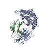
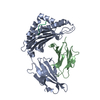
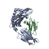
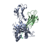
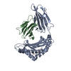
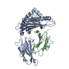
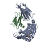
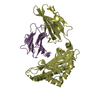
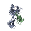
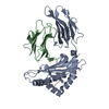
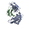
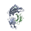
| Title | Phosphorylation of self-peptides alters Human Leukocyte Antigen Class I-restricted antigen presentation and generates tumor specific epitopes | ||||||
 Components Components |
| ||||||
 Keywords Keywords | IMMUNE SYSTEM / PHOSPHORYLATION / Glycoprotein / Immune response / Membrane / MHC I / Phosphoprotein / Polymorphism / Transmembrane / Ubl conjugation / Disease mutation / Immunoglobulin domain / Pyrrolidone carboxylic acid / Secreted / cancer / TCR / self-epitope | ||||||
| Function / homology |  Function and homology information Function and homology informationcellular response to endothelin / negative regulation of long-chain fatty acid import across plasma membrane / IRS-related events triggered by IGF1R / positive regulation of fatty acid beta-oxidation / positive regulation of glucose metabolic process / Signaling by Erythropoietin / Erythropoietin activates STAT5 / phosphatidylinositol 3-kinase activator activity / IRS-mediated signalling / Erythropoietin activates Phospholipase C gamma (PLCG) ...cellular response to endothelin / negative regulation of long-chain fatty acid import across plasma membrane / IRS-related events triggered by IGF1R / positive regulation of fatty acid beta-oxidation / positive regulation of glucose metabolic process / Signaling by Erythropoietin / Erythropoietin activates STAT5 / phosphatidylinositol 3-kinase activator activity / IRS-mediated signalling / Erythropoietin activates Phospholipase C gamma (PLCG) / transmembrane receptor protein tyrosine kinase adaptor activity / Signaling by Leptin / PI3K/AKT activation / positive regulation of memory T cell activation / T cell mediated cytotoxicity directed against tumor cell target / negative regulation of B cell apoptotic process / positive regulation of CD8-positive, alpha-beta T cell activation / CD8-positive, alpha-beta T cell activation / positive regulation of CD8-positive, alpha-beta T cell proliferation / Erythropoietin activates Phosphoinositide-3-kinase (PI3K) / IRS activation / antigen processing and presentation of endogenous peptide antigen via MHC class I via ER pathway, TAP-dependent / TAP complex binding / antigen processing and presentation of exogenous peptide antigen via MHC class I / Golgi medial cisterna / lipid homeostasis / CD8 receptor binding / protection from natural killer cell mediated cytotoxicity / regulation of lipid metabolic process / RET signaling / endoplasmic reticulum exit site / TAP binding / PI3K Cascade / beta-2-microglobulin binding / positive regulation of glycogen biosynthetic process / SOS-mediated signalling / Signal attenuation / detection of bacterium / antigen processing and presentation of endogenous peptide antigen via MHC class Ib / antigen processing and presentation of endogenous peptide antigen via MHC class I via ER pathway, TAP-independent / Growth hormone receptor signaling / response to glucose / phosphatidylinositol 3-kinase binding / Erythropoietin activates RAS / T cell receptor binding / positive regulation of B cell proliferation / insulin-like growth factor receptor signaling pathway / 14-3-3 protein binding / signaling adaptor activity / Interleukin-7 signaling / early endosome lumen / Nef mediated downregulation of MHC class I complex cell surface expression / DAP12 interactions / positive regulation of D-glucose import across plasma membrane / T cell mediated cytotoxicity / Endosomal/Vacuolar pathway / insulin receptor binding / Antigen Presentation: Folding, assembly and peptide loading of class I MHC / lumenal side of endoplasmic reticulum membrane / regulation of iron ion transport / cellular response to glucose stimulus / cellular response to iron(III) ion / antigen processing and presentation of exogenous protein antigen via MHC class Ib, TAP-dependent / negative regulation of iron ion transport / negative regulation of forebrain neuron differentiation / regulation of erythrocyte differentiation / peptide antigen assembly with MHC class I protein complex / ER to Golgi transport vesicle membrane / response to molecule of bacterial origin / HFE-transferrin receptor complex / MHC class I peptide loading complex / transferrin transport / negative regulation of receptor-mediated endocytosis / cellular response to iron ion / positive regulation of T cell cytokine production / antigen processing and presentation of endogenous peptide antigen via MHC class I / MHC class I protein complex / peptide antigen assembly with MHC class II protein complex / negative regulation of neurogenesis / multicellular organismal-level iron ion homeostasis / cellular response to nicotine / MHC class II protein complex / positive regulation of receptor-mediated endocytosis / positive regulation of T cell mediated cytotoxicity / glucose metabolic process / negative regulation of epithelial cell proliferation / specific granule lumen / antigen processing and presentation of exogenous peptide antigen via MHC class II / positive regulation of immune response / peptide antigen binding / phagocytic vesicle membrane / recycling endosome membrane / positive regulation of T cell activation / Interferon gamma signaling / Immunoregulatory interactions between a Lymphoid and a non-Lymphoid cell / cellular response to insulin stimulus / Constitutive Signaling by Aberrant PI3K in Cancer / positive regulation of insulin secretion / positive regulation of type II interferon production / Interferon alpha/beta signaling Similarity search - Function | ||||||
| Biological species |  Homo sapiens (human) Homo sapiens (human) | ||||||
| Method |  X-RAY DIFFRACTION / MOLECULAR REPLACEMENT / Resolution: 1.927 Å X-RAY DIFFRACTION / MOLECULAR REPLACEMENT / Resolution: 1.927 Å | ||||||
 Authors Authors | Petersen, J. / Rossjohn, J. | ||||||
 Citation Citation | Journal: Proc.Natl.Acad.Sci.Usa / Year: 2009 Title: Phosphorylated self-peptides alter human leukocyte antigen class I-restricted antigen presentation and generate tumor-specific epitopes Authors: Petersen, J. / Wurzbacher, S.J. / Williamson, N.A. / Ramarathinam, S.H. / Reid, H.H. / Nair, A.K. / Zhao, A.Y. / Nastovska, R. / Rudge, G. / Rossjohn, J. / Purcell, A.W. | ||||||
| History |
|
- Structure visualization
| Structure viewer | Molecule: MolmilJmol/JSmol |
|---|
- Downloads & links
-Download
| PDBx/mmCIF format | 3fqw.cif.gz | 177.9 KB | Display | PDBx/mmCIF format |
|---|---|---|---|---|
| PDB format | pdb3fqw.ent.gz | 140.2 KB | Display | PDB format |
| PDBx/mmJSON format | 3fqw.json.gz | Tree view | PDBx/mmJSON format | |
| Others |  Other downloads Other downloads |
-Validation report
| Arichive directory | https://data.pdbj.org/pub/pdb/validation_reports/fq/3fqwftp://data.pdbj.org/pub/pdb/validation_reports/fq/3fqw | HTTPS FTP |
|---|
-Related structure data
| Related structure data |  3fqnC  3fqrC  3fqtC  3fquC  3fqxC  2vllS C: citing same article ( S: Starting model for refinement |
|---|---|
| Similar structure data |


-Links
 PDBj
PDBj


- Assembly
| Deposited unit | 
| ||||||||
|---|---|---|---|---|---|---|---|---|---|
| 1 |
| ||||||||
| Unit cell |
|
-Components
-Protein , 2 types, 2 molecules AB
| #1: Protein | Mass: 31854.203 Da / Num. of mol.: 1 Fragment: EXTRACELLULAR DOMAINS ALPHA1, ALPHA2, ALPHA3, UNP residues 25-299 Source method: isolated from a genetically manipulated source Source: (gene. exp.) Homo sapiens (human) / Gene: HLA-A, HLAA / Plasmid: pET 30 / Production host:  |
|---|---|
| #2: Protein | Mass: 11635.002 Da / Num. of mol.: 1 Source method: isolated from a genetically manipulated source Source: (gene. exp.) Homo sapiens (human) / Gene: B2M / Plasmid: pET 30 / Production host: |
-Protein/peptide , 1 types, 1 molecules C
| #3: Protein/peptide | Mass: 873.974 Da / Num. of mol.: 1 / Source method: obtained synthetically / Details: synthetic peptide / References: UniProt: Q9Y4H2*PLUS |
|---|
-Non-polymers , 3 types, 343 molecules 


| #4: Chemical | ChemComp-CD /  Mass: 112.411 Da / Num. of mol.: 5 / Source method: obtained synthetically / Formula: Cd Mass: 112.411 Da / Num. of mol.: 5 / Source method: obtained synthetically / Formula: Cd#5: Chemical | ChemComp-CO / |  Mass: 58.933 Da / Num. of mol.: 1 / Source method: obtained synthetically / Formula: Co Mass: 58.933 Da / Num. of mol.: 1 / Source method: obtained synthetically / Formula: Co#6: Water | ChemComp-HOH / | Mass: 18.015 Da / Num. of mol.: 337 / Source method: isolated from a natural source / Formula: H2O |
|---|
-Details
| Has protein modification | Y |
|---|
-Experimental details
-Experiment
| Experiment | Method: X-RAY DIFFRACTION / Number of used crystals: 1 |
|---|
- Sample preparation
| Crystal | Density Matthews: 2.99 Å3/Da / Density % sol: 58.81 % / Mosaicity: 0.456 ° |
|---|---|
| Crystal grow | Temperature: 293 K / Method: vapor diffusion, hanging drop / pH: 7.4 Details: 13% PEG3350, 10mM CdCl2, 10mM CoCl2, 0.1M NaCl, pH7.4, temperature 293K, VAPOR DIFFUSION, HANGING DROP |
-Data collection
| Diffraction | Mean temperature: 100 K |
|---|---|
| Diffraction source | Source: ROTATING ANODE / Type: RIGAKU RUH3R / Wavelength: 1.5418 Å |
| Detector | Type: RIGAKU RAXIS IV++ / Detector: IMAGE PLATE / Date: May 8, 2008 / Details: osmic mirrors |
| Radiation | Protocol: SINGLE WAVELENGTH / Monochromatic (M) / Laue (L): M / Scattering type: x-ray |
| Radiation wavelength | Wavelength: 1.5418 Å / Relative weight: 1 |
| Reflection | Resolution: 1.927→50 Å / Num. obs: 38037 / % possible obs: 93.1 % / Redundancy: 4.7 % / Rmerge(I) obs: 0.056 / Χ2: 1.014 / Net I/σ(I): 22.144 |
| Reflection shell | Resolution: 1.93→2 Å / Redundancy: 3.9 % / Rmerge(I) obs: 0.328 / Mean I/σ(I) obs: 3.7 / Num. unique all: 3049 / Χ2: 0.992 / % possible all: 75.9 |
- Processing
| Software |
| ||||||||||||||||||||||||||||||||||||||||||||||||||||||||||||||||||||||||||||||||||||||||||||||||||||
|---|---|---|---|---|---|---|---|---|---|---|---|---|---|---|---|---|---|---|---|---|---|---|---|---|---|---|---|---|---|---|---|---|---|---|---|---|---|---|---|---|---|---|---|---|---|---|---|---|---|---|---|---|---|---|---|---|---|---|---|---|---|---|---|---|---|---|---|---|---|---|---|---|---|---|---|---|---|---|---|---|---|---|---|---|---|---|---|---|---|---|---|---|---|---|---|---|---|---|---|---|---|
| Refinement | Method to determine structure: MOLECULAR REPLACEMENT Starting model: PDB ENTRY 2VLL Resolution: 1.927→23.655 Å / Occupancy max: 1 / Occupancy min: 0.5 / FOM work R set: 0.873 / SU ML: 0.23 / Cross valid method: THROUGHOUT / σ(F): 1.34
| ||||||||||||||||||||||||||||||||||||||||||||||||||||||||||||||||||||||||||||||||||||||||||||||||||||
| Solvent computation | Shrinkage radii: 0.9 Å / VDW probe radii: 1.11 Å / Solvent model: FLAT BULK SOLVENT MODEL / Bsol: 42.378 Å2 / ksol: 0.363 e/Å3 | ||||||||||||||||||||||||||||||||||||||||||||||||||||||||||||||||||||||||||||||||||||||||||||||||||||
| Displacement parameters | Biso max: 74.93 Å2 / Biso mean: 28.227 Å2 / Biso min: 8.11 Å2
| ||||||||||||||||||||||||||||||||||||||||||||||||||||||||||||||||||||||||||||||||||||||||||||||||||||
| Refinement step | Cycle: LAST / Resolution: 1.927→23.655 Å
| ||||||||||||||||||||||||||||||||||||||||||||||||||||||||||||||||||||||||||||||||||||||||||||||||||||
| Refine LS restraints |
| ||||||||||||||||||||||||||||||||||||||||||||||||||||||||||||||||||||||||||||||||||||||||||||||||||||
| LS refinement shell | Refine-ID: X-RAY DIFFRACTION / Total num. of bins used: 13
| ||||||||||||||||||||||||||||||||||||||||||||||||||||||||||||||||||||||||||||||||||||||||||||||||||||
| Refinement TLS params. | Method: refined / Refine-ID: X-RAY DIFFRACTION
| ||||||||||||||||||||||||||||||||||||||||||||||||||||||||||||||||||||||||||||||||||||||||||||||||||||
| Refinement TLS group |
|
