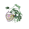
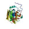
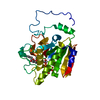
[English] 日本語
Yorodumi
- PDB-3fde: Mouse UHRF1 SRA domain bound with hemi-methylated CpG DNA, crysta... -

+
Open data


ID or keywords:

Loading...

-
Basic information

Entry
Database: PDB / ID: 3fde
TitleMouse UHRF1 SRA domain bound with hemi-methylated CpG DNA, crystal structure in space group C222(1) at 1.4 A resolution
Components
  • 5'-D(*CP*CP*AP*TP*GP*(5CM)P*GP*CP*TP*GP*AP*C)-3'
  • 5'-D(*GP*TP*CP*AP*GP*CP*GP*CP*AP*TP*GP*G)-3'
  • E3 ubiquitin-protein ligase UHRF1
KeywordsLIGASE / SRA domain / base flipping / DNA CpG methylation / Cell cycle / Developmental protein / DNA damage / DNA repair / DNA-binding / Metal-binding / Nucleus / Phosphoprotein / Transcription / Transcription regulation / Ubl conjugation / Ubl conjugation pathway / Zinc / Zinc-finger
Function / homology
Function and homology information


histone H3 ubiquitin ligase activity / hemi-methylated DNA-binding / negative regulation of gene expression via chromosomal CpG island methylation / regulation of epithelial cell proliferation / methyl-CpG binding / mitotic spindle assembly / protein autoubiquitination / heterochromatin / cis-regulatory region sequence-specific DNA binding / heterochromatin formation ...histone H3 ubiquitin ligase activity / hemi-methylated DNA-binding / negative regulation of gene expression via chromosomal CpG island methylation / regulation of epithelial cell proliferation / methyl-CpG binding / mitotic spindle assembly / protein autoubiquitination / heterochromatin / cis-regulatory region sequence-specific DNA binding / heterochromatin formation / methylated histone binding / positive regulation of protein metabolic process / replication fork / euchromatin / RING-type E3 ubiquitin transferase / nuclear matrix / ubiquitin-protein transferase activity / ubiquitin protein ligase activity / histone binding / ubiquitin-dependent protein catabolic process / nucleic acid binding / DNA repair / chromatin / negative regulation of transcription by RNA polymerase II / zinc ion binding / nucleoplasm / identical protein binding / nucleus / cytosol
Similarity search - Function
SRA-YDG / PUA domain-like / : / UHRF1, tandem tudor domain / UHRF1/2-like / Tandem tudor domain within UHRF1 / SRA-YDG / SRA-YDG superfamily / SAD/SRA domain / YDG domain profile. ...SRA-YDG / PUA domain-like / : / UHRF1, tandem tudor domain / UHRF1/2-like / Tandem tudor domain within UHRF1 / SRA-YDG / SRA-YDG superfamily / SAD/SRA domain / YDG domain profile. / SET and RING finger associated domain. Domain of unknown function in SET domain containing proteins and in Deinococcus radiodurans DRA1533. / PUA-like superfamily / Zinc finger, RING-type, conserved site / Zinc finger RING-type signature. / PHD-finger / Ring finger / Zinc finger PHD-type signature. / Zinc finger PHD-type profile. / Zinc finger, PHD-finger / Zinc finger, PHD-type / PHD zinc finger / Zinc finger RING-type profile. / Zinc finger, RING-type / Zinc finger, FYVE/PHD-type / Ubiquitin family / Ubiquitin homologues / Ubiquitin-like domain / Ubiquitin domain profile. / Ubiquitin-like domain superfamily / Zinc finger, RING/FYVE/PHD-type / Roll / Mainly Beta
Similarity search - Domain/homology
Unknown ligand / DNA / DNA (> 10) / E3 ubiquitin-protein ligase UHRF1
Similarity search - Component
Biological speciesMus musculus (house mouse)
MethodX-RAY DIFFRACTION / SYNCHROTRON / MOLECULAR REPLACEMENT / Resolution: 1.41 Å
AuthorsHashimoto, H. / Horton, J.R. / Zhang, X. / Cheng, X.
Citation
Journal: Epigenetics / Year: 2009
Title: UHRF1, a modular multi-domain protein, regulates replication-coupled crosstalk between DNA methylation and histone modifications.
Authors: Hashimoto, H. / Horton, J.R. / Zhang, X. / Cheng, X.
#1: Journal: Nature / Year: 2008
Title: The SRA domain of UHRF1 flips 5-methylcytosine out of the DNA helix.
Authors: Hashimoto, H. / Horton, J.R. / Zhang, X. / Bostick, M. / Jacobsen, S.E. / Cheng, X.
History
DepositionNov 25, 2008Deposition site: RCSB / Processing site: RCSB
Revision 1.0Jan 6, 2009Provider: repository / Type: Initial release
Revision 1.1Jul 13, 2011Group: Version format compliance
Revision 1.2Dec 27, 2023Group: Advisory / Data collection ...Advisory / Data collection / Database references / Derived calculations
Category: chem_comp_atom / chem_comp_bond ...chem_comp_atom / chem_comp_bond / database_2 / pdbx_struct_conn_angle / pdbx_validate_polymer_linkage / struct_conn / struct_ref_seq / struct_ref_seq_dif / struct_site
Item: _database_2.pdbx_DOI / _database_2.pdbx_database_accession ..._database_2.pdbx_DOI / _database_2.pdbx_database_accession / _pdbx_struct_conn_angle.ptnr1_auth_asym_id / _pdbx_struct_conn_angle.ptnr1_auth_comp_id / _pdbx_struct_conn_angle.ptnr1_auth_seq_id / _pdbx_struct_conn_angle.ptnr1_label_asym_id / _pdbx_struct_conn_angle.ptnr1_label_atom_id / _pdbx_struct_conn_angle.ptnr1_label_comp_id / _pdbx_struct_conn_angle.ptnr1_label_seq_id / _pdbx_struct_conn_angle.ptnr3_auth_asym_id / _pdbx_struct_conn_angle.ptnr3_auth_comp_id / _pdbx_struct_conn_angle.ptnr3_auth_seq_id / _pdbx_struct_conn_angle.ptnr3_label_asym_id / _pdbx_struct_conn_angle.ptnr3_label_atom_id / _pdbx_struct_conn_angle.ptnr3_label_comp_id / _pdbx_struct_conn_angle.ptnr3_label_seq_id / _pdbx_struct_conn_angle.value / _pdbx_validate_polymer_linkage.dist / _pdbx_validate_polymer_linkage.label_alt_id_2 / _struct_conn.pdbx_dist_value / _struct_conn.pdbx_leaving_atom_flag / _struct_conn.ptnr1_auth_asym_id / _struct_conn.ptnr1_auth_comp_id / _struct_conn.ptnr1_auth_seq_id / _struct_conn.ptnr1_label_asym_id / _struct_conn.ptnr1_label_atom_id / _struct_conn.ptnr1_label_comp_id / _struct_conn.ptnr1_label_seq_id / _struct_conn.ptnr2_auth_asym_id / _struct_conn.ptnr2_auth_comp_id / _struct_conn.ptnr2_auth_seq_id / _struct_conn.ptnr2_label_asym_id / _struct_conn.ptnr2_label_atom_id / _struct_conn.ptnr2_label_comp_id / _struct_ref_seq.db_align_beg / _struct_ref_seq.db_align_end / _struct_ref_seq_dif.details / _struct_site.pdbx_auth_asym_id / _struct_site.pdbx_auth_comp_id / _struct_site.pdbx_auth_seq_id

-
Structure visualization

Structure viewerMolecule:
MolmilJmol/JSmol

Downloads & links

-
Assembly

Deposited unit
A: E3 ubiquitin-protein ligase UHRF1
B: E3 ubiquitin-protein ligase UHRF1
D: 5'-D(*CP*CP*AP*TP*GP*(5CM)P*GP*CP*TP*GP*AP*C)-3'
E: 5'-D(*GP*TP*CP*AP*GP*CP*GP*CP*AP*TP*GP*G)-3'
C: 5'-D(*CP*CP*AP*TP*GP*(5CM)P*GP*CP*TP*GP*AP*C)-3'
F: 5'-D(*GP*TP*CP*AP*GP*CP*GP*CP*AP*TP*GP*G)-3'
hetero molecules


Theoretical massNumber of molelcules
Total (without water)63,97141
Polymers62,5136
Non-polymers1,45735
Water12,250680
1
A: E3 ubiquitin-protein ligase UHRF1
D: 5'-D(*CP*CP*AP*TP*GP*(5CM)P*GP*CP*TP*GP*AP*C)-3'
E: 5'-D(*GP*TP*CP*AP*GP*CP*GP*CP*AP*TP*GP*G)-3'
hetero molecules


Theoretical massNumber of molelcules
Total (without water)31,98518
Polymers31,2573
Non-polymers72915
Water543
TypeNameSymmetry operationNumber
identity operation1_555x,y,z1
Buried area3180 Å2
ΔGint-15.4 kcal/mol
Surface area13640 Å2
MethodPISA
2
B: E3 ubiquitin-protein ligase UHRF1
C: 5'-D(*CP*CP*AP*TP*GP*(5CM)P*GP*CP*TP*GP*AP*C)-3'
F: 5'-D(*GP*TP*CP*AP*GP*CP*GP*CP*AP*TP*GP*G)-3'
hetero molecules


Theoretical massNumber of molelcules
Total (without water)31,98523
Polymers31,2573
Non-polymers72920
Water543
TypeNameSymmetry operationNumber
identity operation1_555x,y,z1
Buried area3030 Å2
ΔGint-15.4 kcal/mol
Surface area13430 Å2
MethodPISA
Unit cell
Length a, b, c (Å)84.678, 103.691, 149.838
Angle α, β, γ (deg.)90.00, 90.00, 90.00
Int Tables number20
Space group name H-MC2221
DetailsAsymmetric unit contains two SRA-DNA complexes.

-
Components

-
Protein , 1 types, 2 molecules AB

#1: Protein E3 ubiquitin-protein ligase UHRF1 / Ubiquitin-like PHD and RING finger domain-containing protein 1 / Ubiquitin-like-containing PHD and ...Ubiquitin-like PHD and RING finger domain-containing protein 1 / Ubiquitin-like-containing PHD and RING finger domains protein 1 / Nuclear zinc finger protein Np95 / Nuclear protein 95


Mass: 23915.711 Da / Num. of mol.: 2 / Fragment: YDG domain: UNP residues 419-628
Source method: isolated from a genetically manipulated source
Details: hexahistidine-SUMO-tagged construct / Source: (gene. exp.) Mus musculus (house mouse) / Gene: Np95, Uhrf1 / Plasmid: pXC666 / Production host: Escherichia coli (E. coli)
References: UniProt: Q8VDF2, Ligases; Forming carbon-nitrogen bonds; Acid-amino-acid ligases (peptide synthases)

-
DNA chain , 2 types, 4 molecules DCEF

#2: DNA chain 5'-D(*CP*CP*AP*TP*GP*(5CM)P*GP*CP*TP*GP*AP*C)-3'


Mass: 3637.395 Da / Num. of mol.: 2 / Source method: obtained synthetically / Details: synthetic DNA oligo
#3: DNA chain 5'-D(*GP*TP*CP*AP*GP*CP*GP*CP*AP*TP*GP*G)-3'


Mass: 3703.416 Da / Num. of mol.: 2 / Source method: obtained synthetically / Details: synthetic DNA oligo

-
Non-polymers , 4 types, 715 molecules

#4: Chemical...
ChemComp-EDO / 1,2-ETHANEDIOL / ETHYLENE GLYCOL / Ethylene glycol


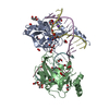
Mass: 62.068 Da / Num. of mol.: 22 / Source method: obtained synthetically / Formula: C2H6O2
#5: Chemical
ChemComp-NA / SODIUM ION


Mass: 22.990 Da / Num. of mol.: 4 / Source method: obtained synthetically / Formula: Na
#6: Chemical
ChemComp-UNL / UNKNOWN LIGAND


Num. of mol.: 9 / Source method: obtained synthetically
#7: Water ChemComp-HOH / water / Water


Mass: 18.015 Da / Num. of mol.: 680 / Source method: isolated from a natural source / Formula: H2O

-
Experimental details

-
Experiment

ExperimentMethod: X-RAY DIFFRACTION / Number of used crystals: 1

-
Sample preparation

CrystalDensity Matthews: 2.63 Å3/Da / Density % sol: 53.24 %
Crystal growTemperature: 277 K / Method: vapor diffusion / pH: 7
Details: 20% PEG 3350, 0.4M NaCl, pH 7.0, VAPOR DIFFUSION, temperature 277K
Components of the solutions
IDNameCrystal-IDSol-ID
1PEG 335011
2NaClSodium chloride11
3PEG 335012
4NaClSodium chloride12

-
Data collection

DiffractionMean temperature: 100 K
Diffraction sourceSource: SYNCHROTRON / Site: APS / Beamline: 22-ID / Wavelength: 1 Å
DetectorType: MARMOSAIC 300 mm CCD / Detector: CCD / Date: Aug 6, 2008
RadiationProtocol: SINGLE WAVELENGTH / Monochromatic (M) / Laue (L): M / Scattering type: x-ray
Radiation wavelengthWavelength: 1 Å / Relative weight: 1
ReflectionResolution: 1.41→32.79 Å / Num. obs: 121098 / % possible obs: 97 % / Observed criterion σ(I): -3 / Redundancy: 5.1 % / Biso Wilson estimate: 12.9 Å2 / Rmerge(I) obs: 0.095 / Net I/σ(I): 10.9
Reflection shellResolution: 1.41→1.45 Å / Redundancy: 1.7 % / Rmerge(I) obs: 0.771 / Mean I/σ(I) obs: 1

-
Processing

Software
NameVersionClassification
HKL-2000data collection
PHASERphasing
REFMAC5.5.0044refinement
HKL-2000data reduction
HKL-2000data scaling
RefinementMethod to determine structure: MOLECULAR REPLACEMENT / Resolution: 1.41→32.79 Å / Cor.coef. Fo:Fc: 0.975 / Cor.coef. Fo:Fc free: 0.962 / SU B: 2.047 / SU ML: 0.035 / Isotropic thermal model: PDB entry 2ZO0 / Cross valid method: THROUGHOUT / ESU R: 0.06 / ESU R Free: 0.057 / Stereochemistry target values: MAXIMUM LIKELIHOOD / Details: HYDROGENS HAVE BEEN ADDED IN THE RIDING POSITIONS
RfactorNum. reflection% reflectionSelection details
Rfree0.18632 6067 5 %RANDOM
Rwork0.14906 ---
obs0.15093 114952 96.07 %-
Solvent computationIon probe radii: 0.8 Å / Shrinkage radii: 0.8 Å / VDW probe radii: 1.4 Å / Solvent model: MASK
Displacement parametersBiso mean: 19.522 Å2
Baniso -1Baniso -2Baniso -3
1-0.03 Å20 Å2-0 Å2
2---0.08 Å20 Å2
3---0.05 Å2
Refine analyze
FreeObs
Luzzati coordinate error0.057 Å0.06 Å
Luzzati sigma a-0.035 Å
Refinement stepCycle: LAST / Resolution: 1.41→32.79 Å
ProteinNucleic acidLigandSolventTotal
Num. atoms3349 1016 114 680 5159
Refine LS restraints
Refine-IDTypeDev idealDev ideal targetNumber
X-RAY DIFFRACTIONr_bond_refined_d0.0220.0214662
X-RAY DIFFRACTIONr_angle_refined_deg2.122.2286454
X-RAY DIFFRACTIONr_dihedral_angle_1_deg5.5635436
X-RAY DIFFRACTIONr_dihedral_angle_2_deg32.39922.528178
X-RAY DIFFRACTIONr_dihedral_angle_3_deg12.58115550
X-RAY DIFFRACTIONr_dihedral_angle_4_deg18.8631540
X-RAY DIFFRACTIONr_chiral_restr0.2050.2651
X-RAY DIFFRACTIONr_gen_planes_refined0.0150.0213326
X-RAY DIFFRACTIONr_mcbond_it2.1891.52117
X-RAY DIFFRACTIONr_mcangle_it3.16123380
X-RAY DIFFRACTIONr_scbond_it4.07532476
X-RAY DIFFRACTIONr_scangle_it5.4944.53035
X-RAY DIFFRACTIONr_rigid_bond_restr2.61334658
X-RAY DIFFRACTIONr_sphericity_free10.0953704
X-RAY DIFFRACTIONr_sphericity_bonded6.56134454
LS refinement shellResolution: 1.41→1.45 Å / Total num. of bins used: 20
RfactorNum. reflection% reflection
Rfree0.453 279 -
Rwork0.481 5243 -
obs-5243 60.41 %

+
About Yorodumi

-
News

-
Feb 9, 2022. New format data for meta-information of EMDB entries

New format data for meta-information of EMDB entries

  • Version 3 of the EMDB header file is now the official format.
  • The previous official version 1.9 will be removed from the archive.

Related info.:EMDB header

External links:wwPDB to switch to version 3 of the EMDB data model

-
Aug 12, 2020. Covid-19 info

Covid-19 info

URL: https://pdbj.org/emnavi/covid19.php

New page: Covid-19 featured information page in EM Navigator.

Related info.:Covid-19 info / Mar 5, 2020. Novel coronavirus structure data

+
Mar 5, 2020. Novel coronavirus structure data

Novel coronavirus structure data

Related info.:Yorodumi Speices / Aug 12, 2020. Covid-19 info

External links:COVID-19 featured content - PDBj / Molecule of the Month (242):Coronavirus Proteases

+
Jan 31, 2019. EMDB accession codes are about to change! (news from PDBe EMDB page)

EMDB accession codes are about to change! (news from PDBe EMDB page)

  • The allocation of 4 digits for EMDB accession codes will soon come to an end. Whilst these codes will remain in use, new EMDB accession codes will include an additional digit and will expand incrementally as the available range of codes is exhausted. The current 4-digit format prefixed with “EMD-” (i.e. EMD-XXXX) will advance to a 5-digit format (i.e. EMD-XXXXX), and so on. It is currently estimated that the 4-digit codes will be depleted around Spring 2019, at which point the 5-digit format will come into force.
  • The EM Navigator/Yorodumi systems omit the EMD- prefix.

Related info.:Q: What is EMD? / ID/Accession-code notation in Yorodumi/EM Navigator

External links:EMDB Accession Codes are Changing Soon! / Contact to PDBj

+
Jul 12, 2017. Major update of PDB

Major update of PDB

  • wwPDB released updated PDB data conforming to the new PDBx/mmCIF dictionary.
  • This is a major update changing the version number from 4 to 5, and with Remediation, in which all the entries are updated.
  • In this update, many items about electron microscopy experimental information are reorganized (e.g. em_software).
  • Now, EM Navigator and Yorodumi are based on the updated data.

External links:wwPDB Remediation / Enriched Model Files Conforming to OneDep Data Standards Now Available in the PDB FTP Archive

-
Yorodumi

Thousand views of thousand structures

  • Yorodumi is a browser for structure data from EMDB, PDB, SASBDB, etc.
  • This page is also the successor to EM Navigator detail page, and also detail information page/front-end page for Omokage search.
  • The word "yorodu" (or yorozu) is an old Japanese word meaning "ten thousand". "mi" (miru) is to see.

Related info.:EMDB / PDB / SASBDB / Comparison of 3 databanks / Yorodumi Search / Aug 31, 2016. New EM Navigator & Yorodumi / Yorodumi Papers / Jmol/JSmol / Function and homology information / Changes in new EM Navigator and Yorodumi

Read more