 Movie
Movie Controller

[English] 日本語
 Yorodumi
- PDB-3f8j: Mouse UHRF1 SRA domain bound with hemi-methylated CpG, crystal st... -
+ Open data

- Basic information
| Entry | Database: PDB / ID: 3f8j | ||||||
|---|---|---|---|---|---|---|---|
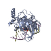
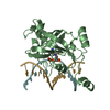
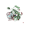
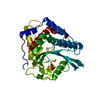
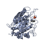
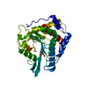
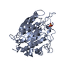
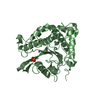
| Title | Mouse UHRF1 SRA domain bound with hemi-methylated CpG, crystal structure in space group C222(1) | ||||||
 Components Components |
| ||||||
 Keywords Keywords | LIGASE/DNA / UHRF1 / SRA / base flipping / 5-methylcytosine / CpG methylation / Cell cycle / Developmental protein / DNA damage / DNA repair / DNA-binding / Ligase / Metal-binding / Nucleus / Phosphoprotein / Transcription / Transcription regulation / Ubl conjugation pathway / Zinc-finger / LIGASE-DNA COMPLEX | ||||||
| Function / homology |  Function and homology information Function and homology informationhistone H3K18 ubiquitin ligase activity / histone H3K14 ubiquitin ligase activity / histone H3 ubiquitin ligase activity / histone H3K23 ubiquitin ligase activity / histone H3 reader activity / chromosomal DNA methylation maintenance following DNA replication / hemi-methylated DNA-binding / regulation of epithelial cell proliferation / methyl-CpG binding / histone H3K9me2/3 reader activity ...histone H3K18 ubiquitin ligase activity / histone H3K14 ubiquitin ligase activity / histone H3 ubiquitin ligase activity / histone H3K23 ubiquitin ligase activity / histone H3 reader activity / chromosomal DNA methylation maintenance following DNA replication / hemi-methylated DNA-binding / regulation of epithelial cell proliferation / methyl-CpG binding / histone H3K9me2/3 reader activity / negative regulation of gene expression via chromosomal CpG island methylation / positive regulation of protein metabolic process / mitotic spindle assembly / protein autoubiquitination / cis-regulatory region sequence-specific DNA binding / heterochromatin / replication fork / euchromatin / RING-type E3 ubiquitin transferase / nuclear matrix / ubiquitin-protein transferase activity / ubiquitin protein ligase activity / heterochromatin formation / histone binding / nucleic acid binding / ubiquitin-dependent protein catabolic process / protein ubiquitination / DNA repair / chromatin / negative regulation of transcription by RNA polymerase II / positive regulation of transcription by RNA polymerase II / zinc ion binding / nucleoplasm / identical protein binding / nucleus / cytosol Similarity search - Function | ||||||
| Biological species |  | ||||||
| Method |  X-RAY DIFFRACTION / SYNCHROTRON / MOLECULAR REPLACEMENT / Resolution: 1.99 Å X-RAY DIFFRACTION / SYNCHROTRON / MOLECULAR REPLACEMENT / Resolution: 1.99 Å | ||||||
 Authors Authors | Hashimoto, H. / Horton, J.R. / Zhang, X. / Cheng, X. | ||||||
 Citation Citation | Journal: Epigenetics / Year: 2009 Title: UHRF1, a modular multi-domain protein, regulates replication-coupled crosstalk between DNA methylation and histone modifications. Authors: Hashimoto, H. / Horton, J.R. / Zhang, X. / Cheng, X. #1: Journal: Nature / Year: 2008Title: The SRA domain of UHRF1 flips 5-methylcytosine out of the DNA helix. Authors: Hashimoto, H. / Horton, J.R. / Zhang, X. / Bostick, M. / Jacobsen, S.E. / Cheng, X. | ||||||
| History |
|
- Structure visualization
| Structure viewer | Molecule: MolmilJmol/JSmol |
|---|
- Downloads & links
-Download
| PDBx/mmCIF format | 3f8j.cif.gz | 77.2 KB | Display | PDBx/mmCIF format |
|---|---|---|---|---|
| PDB format | pdb3f8j.ent.gz | 53 KB | Display | PDB format |
| PDBx/mmJSON format | 3f8j.json.gz | Tree view | PDBx/mmJSON format | |
| Others |  Other downloads Other downloads |
-Validation report
| Arichive directory | https://data.pdbj.org/pub/pdb/validation_reports/f8/3f8jftp://data.pdbj.org/pub/pdb/validation_reports/f8/3f8j | HTTPS FTP |
|---|
-Related structure data
| Related structure data |  3f8iC  3fdeC 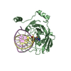 2zo0S S: Starting model for refinement C: citing same article ( |
|---|---|
| Similar structure data |


-Links
 PDBj
PDBj


- Assembly
| Deposited unit | 
| ||||||||
|---|---|---|---|---|---|---|---|---|---|
| 1 |
| ||||||||
| Unit cell |
| ||||||||
| Details | One SRA-DNA complex per asymmetric unit |
-Components
| #1: Protein | Mass: 23858.656 Da / Num. of mol.: 1 / Fragment: YDG domain: UNP residues 417-628 Source method: isolated from a genetically manipulated source Details: hexahistidine-SUMO-tagged construct / Source: (gene. exp.)  | ||||
|---|---|---|---|---|---|
| #2: DNA chain | Mass: 3637.395 Da / Num. of mol.: 1 / Source method: obtained synthetically / Details: Synthetic DNA oligo | ||||
| #3: DNA chain | Mass: 3703.416 Da / Num. of mol.: 1 / Source method: obtained synthetically / Details: Synthetic DNA oligo | ||||
| #4: Chemical | ChemComp-GOL /   Mass: 92.094 Da / Num. of mol.: 9 / Source method: obtained synthetically / Formula: C3H8O3 Mass: 92.094 Da / Num. of mol.: 9 / Source method: obtained synthetically / Formula: C3H8O3#5: Water | ChemComp-HOH / |  Mass: 18.015 Da / Num. of mol.: 161 / Source method: isolated from a natural source / Formula: H2O Mass: 18.015 Da / Num. of mol.: 161 / Source method: isolated from a natural source / Formula: H2OSequence details | AUTHORS USED NATIVE SEQUENCE ILE VAL FOR RESIDUES 417 AND 418 IN THIS ENTRY (WITHOUT ANY EXPRESSION | |
-Experimental details
-Experiment
| Experiment | Method: X-RAY DIFFRACTION / Number of used crystals: 1 |
|---|
- Sample preparation
| Crystal | Density Matthews: 2.68 Å3/Da / Density % sol: 54.1 % | ||||||||||||||||||||
|---|---|---|---|---|---|---|---|---|---|---|---|---|---|---|---|---|---|---|---|---|---|
| Crystal grow | Temperature: 277 K / pH: 7 Details: 20% PEG 3350, 0.4 M NaCl, pH 7.0, VAPOR DIFFUSION, temperature 277K | ||||||||||||||||||||
| Components of the solutions |
|
-Data collection
| Diffraction | Mean temperature: 100 K |
|---|---|
| Diffraction source | Source: SYNCHROTRON / Site: APS  / Beamline: 22-ID / Wavelength: 1 / Beamline: 22-ID / Wavelength: 1 |
| Detector | Type: MARMOSAIC 300 mm CCD / Detector: CCD / Date: Jun 14, 2008 |
| Radiation | Protocol: SINGLE WAVELENGTH / Monochromatic (M) / Laue (L): M / Scattering type: x-ray |
| Radiation wavelength | Wavelength: 1 Å / Relative weight: 1 |
| Reflection | Resolution: 1.99→29.91 Å / Num. obs: 23488 / % possible obs: 99.8 % / Observed criterion σ(I): -3 / Redundancy: 9.6 % / Biso Wilson estimate: 21.8 Å2 / Rmerge(I) obs: 0.102 / Net I/σ(I): 9 |
| Reflection shell | Resolution: 1.99→2.06 Å / Redundancy: 6.5 % / Rmerge(I) obs: 0.568 / Mean I/σ(I) obs: 3.6 / % possible all: 99.1 |
- Processing
| Software |
| ||||||||||||||||||||||||||||||||||||||||||||||||||||||||||||||||||||||||||||||||
|---|---|---|---|---|---|---|---|---|---|---|---|---|---|---|---|---|---|---|---|---|---|---|---|---|---|---|---|---|---|---|---|---|---|---|---|---|---|---|---|---|---|---|---|---|---|---|---|---|---|---|---|---|---|---|---|---|---|---|---|---|---|---|---|---|---|---|---|---|---|---|---|---|---|---|---|---|---|---|---|---|---|
| Refinement | Method to determine structure: MOLECULAR REPLACEMENT Starting model: PDB ENTRY 2ZO0 Resolution: 1.99→29.91 Å / Rfactor Rfree error: 0.007 / Data cutoff high absF: 412547.2 / Data cutoff low absF: 0 / Cross valid method: THROUGHOUT / σ(F): 0 / Stereochemistry target values: ENGH & HUBER
| ||||||||||||||||||||||||||||||||||||||||||||||||||||||||||||||||||||||||||||||||
| Solvent computation | Bsol: 46.64 Å2 / ksol: 0.4 e/Å3 | ||||||||||||||||||||||||||||||||||||||||||||||||||||||||||||||||||||||||||||||||
| Displacement parameters | Biso mean: 28.8 Å2
| ||||||||||||||||||||||||||||||||||||||||||||||||||||||||||||||||||||||||||||||||
| Refine analyze |
| ||||||||||||||||||||||||||||||||||||||||||||||||||||||||||||||||||||||||||||||||
| Refinement step | Cycle: LAST / Resolution: 1.99→29.91 Å
| ||||||||||||||||||||||||||||||||||||||||||||||||||||||||||||||||||||||||||||||||
| Refine LS restraints |
| ||||||||||||||||||||||||||||||||||||||||||||||||||||||||||||||||||||||||||||||||
| LS refinement shell | Resolution: 1.99→2.06 Å / Rfactor Rfree error: 0.029 / Total num. of bins used: 10
| ||||||||||||||||||||||||||||||||||||||||||||||||||||||||||||||||||||||||||||||||
| Xplor file |
|
