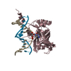
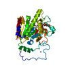
[English] 日本語
Yorodumi
- PDB-2zo0: mouse NP95 SRA domain DNA specific complex 1 -

+
Open data


ID or keywords:

Loading...

-
Basic information

Entry
Database: PDB / ID: 2zo0
Titlemouse NP95 SRA domain DNA specific complex 1
Components
  • DNA (5'-D(*DGP*DTP*DCP*DAP*DGP*(5CM)P*DGP*DCP*DAP*DAP*DTP*DGP*DG)-3')
  • DNA (5'-D(*DTP*DCP*DCP*DAP*DTP*DGP*DCP*DGP*DCP*DTP*DGP*DAP*DC)-3')
  • E3 ubiquitin-protein ligase UHRF1
KeywordsLIGASE/DNA / base flipping / Cell cycle / Developmental protein / DNA damage / DNA repair / DNA-binding / Ligase / Metal-binding / Nucleus / Phosphoprotein / Transcription / Transcription regulation / Ubl conjugation / Ubl conjugation pathway / Zinc / Zinc-finger / LIGASE-DNA COMPLEX
Function / homology
Function and homology information


histone H3 ubiquitin ligase activity / hemi-methylated DNA-binding / regulation of epithelial cell proliferation / methyl-CpG binding / negative regulation of gene expression via chromosomal CpG island methylation / mitotic spindle assembly / protein autoubiquitination / cis-regulatory region sequence-specific DNA binding / heterochromatin / heterochromatin formation ...histone H3 ubiquitin ligase activity / hemi-methylated DNA-binding / regulation of epithelial cell proliferation / methyl-CpG binding / negative regulation of gene expression via chromosomal CpG island methylation / mitotic spindle assembly / protein autoubiquitination / cis-regulatory region sequence-specific DNA binding / heterochromatin / heterochromatin formation / methylated histone binding / positive regulation of protein metabolic process / replication fork / euchromatin / RING-type E3 ubiquitin transferase / nuclear matrix / ubiquitin protein ligase activity / histone binding / ubiquitin-dependent protein catabolic process / protein ubiquitination / DNA repair / chromatin / negative regulation of transcription by RNA polymerase II / zinc ion binding / nucleoplasm / identical protein binding / nucleus / cytosol
Similarity search - Function
SRA-YDG / PUA domain-like / : / UHRF1, tandem tudor domain / UHRF1/2-like / Tandem tudor domain within UHRF1 / SRA-YDG / SRA-YDG superfamily / SAD/SRA domain / YDG domain profile. ...SRA-YDG / PUA domain-like / : / UHRF1, tandem tudor domain / UHRF1/2-like / Tandem tudor domain within UHRF1 / SRA-YDG / SRA-YDG superfamily / SAD/SRA domain / YDG domain profile. / SET and RING finger associated domain. Domain of unknown function in SET domain containing proteins and in Deinococcus radiodurans DRA1533. / PUA-like superfamily / Zinc finger, RING-type, conserved site / Zinc finger RING-type signature. / PHD-finger / Ring finger / Zinc finger PHD-type signature. / Zinc finger PHD-type profile. / Zinc finger, PHD-finger / Zinc finger, PHD-type / PHD zinc finger / Zinc finger RING-type profile. / Zinc finger, RING-type / Zinc finger, FYVE/PHD-type / Ubiquitin family / Ubiquitin homologues / Ubiquitin domain profile. / Ubiquitin-like domain / Zinc finger, RING/FYVE/PHD-type / Ubiquitin-like domain superfamily / Roll / Mainly Beta
Similarity search - Domain/homology
DNA / DNA (> 10) / E3 ubiquitin-protein ligase UHRF1
Similarity search - Component
Biological speciesMus musculus (house mouse)
MethodX-RAY DIFFRACTION / SYNCHROTRON / MOLECULAR REPLACEMENT / Resolution: 2.19 Å
AuthorsHashimoto, H. / Horton, J.R. / Cheng, X.
CitationJournal: Nature / Year: 2008
Title: The SRA domain of UHRF1 flips 5-methylcytosine out of the DNA helix
Authors: Hashimoto, H. / Horton, J.R. / Zhang, X. / Bostick, M. / Jacobsen, S.E. / Cheng, X.
History
DepositionMay 5, 2008Deposition site: PDBJ / Processing site: PDBJ
Revision 1.0Sep 9, 2008Provider: repository / Type: Initial release
Revision 1.1Jul 13, 2011Group: Version format compliance
Revision 1.2Nov 1, 2023Group: Data collection / Database references ...Data collection / Database references / Derived calculations / Refinement description
Category: chem_comp_atom / chem_comp_bond ...chem_comp_atom / chem_comp_bond / database_2 / pdbx_initial_refinement_model / struct_conn / struct_ref_seq / struct_ref_seq_dif
Item: _database_2.pdbx_DOI / _database_2.pdbx_database_accession ..._database_2.pdbx_DOI / _database_2.pdbx_database_accession / _struct_conn.pdbx_leaving_atom_flag / _struct_ref_seq.db_align_beg / _struct_ref_seq.db_align_end / _struct_ref_seq_dif.details

-
Structure visualization

Structure viewerMolecule:
MolmilJmol/JSmol

Downloads & links

-
Assembly

Deposited unit
B: E3 ubiquitin-protein ligase UHRF1
D: DNA (5'-D(*DTP*DCP*DCP*DAP*DTP*DGP*DCP*DGP*DCP*DTP*DGP*DAP*DC)-3')
E: DNA (5'-D(*DGP*DTP*DCP*DAP*DGP*(5CM)P*DGP*DCP*DAP*DAP*DTP*DGP*DG)-3')


Theoretical massNumber of molelcules
Total (without water)31,8743
Polymers31,8743
Non-polymers00
Water75742
1


  • Idetical with deposited unit
  • defined by author&software
TypeNameSymmetry operationNumber
identity operation1_555x,y,z1
Buried area3990 Å2
ΔGint-12 kcal/mol
Surface area13500 Å2
MethodPISA
2
B: E3 ubiquitin-protein ligase UHRF1


Theoretical massNumber of molelcules
Total (without water)23,9161
Polymers23,9161
Non-polymers00
Water181
TypeNameSymmetry operationNumber
identity operation1_555x,y,z1
MethodPISA
3
D: DNA (5'-D(*DTP*DCP*DCP*DAP*DTP*DGP*DCP*DGP*DCP*DTP*DGP*DAP*DC)-3')
E: DNA (5'-D(*DGP*DTP*DCP*DAP*DGP*(5CM)P*DGP*DCP*DAP*DAP*DTP*DGP*DG)-3')


Theoretical massNumber of molelcules
Total (without water)7,9582
Polymers7,9582
Non-polymers00
Water362
TypeNameSymmetry operationNumber
identity operation1_555x,y,z1
Buried area1260 Å2
ΔGint-6 kcal/mol
Surface area5320 Å2
MethodPISA
Unit cell
Length a, b, c (Å)61.982, 69.008, 92.956
Angle α, β, γ (deg.)90.00, 90.00, 90.00
Int Tables number19
Space group name H-MP212121

-
Components

#1: Protein E3 ubiquitin-protein ligase UHRF1 / Ubiquitin-like PHD and RING finger domain-containing protein 1 / Ubiquitin-like-containing PHD and ...Ubiquitin-like PHD and RING finger domain-containing protein 1 / Ubiquitin-like-containing PHD and RING finger domains protein 1 / Nuclear zinc finger protein Np95 / Nuclear protein 95


Mass: 23915.711 Da / Num. of mol.: 1 / Fragment: SRA domain, residues 419-628
Source method: isolated from a genetically manipulated source
Source: (gene. exp.) Mus musculus (house mouse) / Gene: Np95 / Plasmid: pXC666 / Production host: Escherichia coli (E. coli) / Strain (production host): BL21(DE3)-Gold Cells
References: UniProt: Q8VDF2, Ligases; Forming carbon-nitrogen bonds; Acid-amino-acid ligases (peptide synthases)
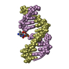
#2: DNA chain DNA (5'-D(*DTP*DCP*DCP*DAP*DTP*DGP*DCP*DGP*DCP*DTP*DGP*DAP*DC)-3')


Mass: 3927.561 Da / Num. of mol.: 1 / Source method: obtained synthetically / Details: 12 base pair DNA duplex
#3: DNA chain DNA (5'-D(*DGP*DTP*DCP*DAP*DGP*(5CM)P*DGP*DCP*DAP*DAP*DTP*DGP*DG)-3')


Mass: 4030.650 Da / Num. of mol.: 1 / Source method: obtained synthetically / Details: 12 base pair DNA duplex
#4: Water ChemComp-HOH / water


Mass: 18.015 Da / Num. of mol.: 42 / Source method: isolated from a natural source / Formula: H2O

-
Experimental details

-
Experiment

ExperimentMethod: X-RAY DIFFRACTION / Number of used crystals: 1

-
Sample preparation

CrystalDensity Matthews: 3.12 Å3/Da / Density % sol: 60.56 %
Crystal growTemperature: 289 K / Method: vapor diffusion, hanging drop / pH: 7
Details: 20% (v/v) polyethylene glycol 3350, 0.4M NaCl, pH 7.0, VAPOR DIFFUSION, HANGING DROP, temperature 289K
Components of the solutions
IDNameCrystal-IDSol-ID
1PEG 335011
2NaCL11
3H2O11
4PEG 335012
5NaCL12
6H2O12

-
Data collection

DiffractionMean temperature: 100 K
Diffraction sourceSource: SYNCHROTRON / Site: APS / Beamline: 22-ID / Wavelength: 1 Å
DetectorType: MAR CCD 165 mm / Detector: CCD / Date: Feb 9, 2008
RadiationProtocol: SINGLE WAVELENGTH / Monochromatic (M) / Laue (L): M / Scattering type: x-ray
Radiation wavelengthWavelength: 1 Å / Relative weight: 1
ReflectionResolution: 2.19→29.4 Å / Num. obs: 19727 / % possible obs: 91.9 % / Observed criterion σ(F): 0 / Observed criterion σ(I): -3 / Redundancy: 10.4 % / Biso Wilson estimate: 20 Å2 / Rmerge(I) obs: 0.073 / Net I/σ(I): 21.8
Reflection shellResolution: 2.19→2.27 Å / Rmerge(I) obs: 0.292 / Mean I/σ(I) obs: 5.4 / Num. unique all: 1343 / % possible all: 63.7

-
Processing

Software
NameVersionClassification
HKL-2000data collection
PHASERphasing
CNS1.2refinement
HKL-2000data reduction
HKL-2000data scaling
RefinementMethod to determine structure: MOLECULAR REPLACEMENT
Starting model: PDB ENTRY 3BI7
Resolution: 2.19→29.4 Å / Rfactor Rfree error: 0.008 / Data cutoff high absF: 708338.01 / Data cutoff low absF: 0 / Isotropic thermal model: RESTRAINED / Cross valid method: THROUGHOUT / σ(F): 0 / Stereochemistry target values: ENGH & HUBER
Details: The density for the nucleotide at position 9 in chain E indicated an extrahelical conformation (E429) as well as an intrahelical conformation (E430). The depositors made a model as if an ...Details: The density for the nucleotide at position 9 in chain E indicated an extrahelical conformation (E429) as well as an intrahelical conformation (E430). The depositors made a model as if an additional nucleotide A10 was inserted.
RfactorNum. reflection% reflectionSelection details
Rfree0.253 897 4.8 %RANDOM
Rwork0.217 ---
obs0.217 18627 88.1 %-
Solvent computationSolvent model: FLAT MODEL / Bsol: 53.074 Å2 / ksol: 0.4 e/Å3
Displacement parametersBiso mean: 71.5 Å2
Baniso -1Baniso -2Baniso -3
1--36.42 Å20 Å20 Å2
2--30.56 Å20 Å2
3---5.86 Å2
Refine analyze
FreeObs
Luzzati coordinate error0.38 Å0.33 Å
Luzzati d res low-35 Å
Luzzati sigma a0.36 Å0.4 Å
Refinement stepCycle: LAST / Resolution: 2.19→29.4 Å
ProteinNucleic acidLigandSolventTotal
Num. atoms1641 519 0 42 2202
Refine LS restraints
Refine-IDTypeDev idealDev ideal target
X-RAY DIFFRACTIONc_bond_d0.006
X-RAY DIFFRACTIONc_bond_d_na
X-RAY DIFFRACTIONc_bond_d_prot
X-RAY DIFFRACTIONc_angle_d
X-RAY DIFFRACTIONc_angle_d_na
X-RAY DIFFRACTIONc_angle_d_prot
X-RAY DIFFRACTIONc_angle_deg1.1
X-RAY DIFFRACTIONc_angle_deg_na
X-RAY DIFFRACTIONc_angle_deg_prot
X-RAY DIFFRACTIONc_dihedral_angle_d22.3
X-RAY DIFFRACTIONc_dihedral_angle_d_na
X-RAY DIFFRACTIONc_dihedral_angle_d_prot
X-RAY DIFFRACTIONc_improper_angle_d0.95
X-RAY DIFFRACTIONc_improper_angle_d_na
X-RAY DIFFRACTIONc_improper_angle_d_prot
X-RAY DIFFRACTIONc_mcbond_it1.31.5
X-RAY DIFFRACTIONc_mcangle_it2.122
X-RAY DIFFRACTIONc_scbond_it1.992
X-RAY DIFFRACTIONc_scangle_it3.052.5
LS refinement shellResolution: 2.19→2.27 Å / Rfactor Rfree error: 0.048 / Total num. of bins used: 10
RfactorNum. reflection% reflection
Rfree0.375 61 5.2 %
Rwork0.367 1114 -
obs-1114 57.2 %

+
About Yorodumi

-
News

-
Feb 9, 2022. New format data for meta-information of EMDB entries

New format data for meta-information of EMDB entries

  • Version 3 of the EMDB header file is now the official format.
  • The previous official version 1.9 will be removed from the archive.

Related info.:EMDB header

External links:wwPDB to switch to version 3 of the EMDB data model

-
Aug 12, 2020. Covid-19 info

Covid-19 info

URL: https://pdbjlvh1.pdbj.org/emnavi/covid19.php

New page: Covid-19 featured information page in EM Navigator.

Related info.:Covid-19 info / Mar 5, 2020. Novel coronavirus structure data

+
Mar 5, 2020. Novel coronavirus structure data

Novel coronavirus structure data

Related info.:Yorodumi Speices / Aug 12, 2020. Covid-19 info

External links:COVID-19 featured content - PDBj / Molecule of the Month (242):Coronavirus Proteases

+
Jan 31, 2019. EMDB accession codes are about to change! (news from PDBe EMDB page)

EMDB accession codes are about to change! (news from PDBe EMDB page)

  • The allocation of 4 digits for EMDB accession codes will soon come to an end. Whilst these codes will remain in use, new EMDB accession codes will include an additional digit and will expand incrementally as the available range of codes is exhausted. The current 4-digit format prefixed with “EMD-” (i.e. EMD-XXXX) will advance to a 5-digit format (i.e. EMD-XXXXX), and so on. It is currently estimated that the 4-digit codes will be depleted around Spring 2019, at which point the 5-digit format will come into force.
  • The EM Navigator/Yorodumi systems omit the EMD- prefix.

Related info.:Q: What is EMD? / ID/Accession-code notation in Yorodumi/EM Navigator

External links:EMDB Accession Codes are Changing Soon! / Contact to PDBj

+
Jul 12, 2017. Major update of PDB

Major update of PDB

  • wwPDB released updated PDB data conforming to the new PDBx/mmCIF dictionary.
  • This is a major update changing the version number from 4 to 5, and with Remediation, in which all the entries are updated.
  • In this update, many items about electron microscopy experimental information are reorganized (e.g. em_software).
  • Now, EM Navigator and Yorodumi are based on the updated data.

External links:wwPDB Remediation / Enriched Model Files Conforming to OneDep Data Standards Now Available in the PDB FTP Archive

-
Yorodumi

Thousand views of thousand structures

  • Yorodumi is a browser for structure data from EMDB, PDB, SASBDB, etc.
  • This page is also the successor to EM Navigator detail page, and also detail information page/front-end page for Omokage search.
  • The word "yorodu" (or yorozu) is an old Japanese word meaning "ten thousand". "mi" (miru) is to see.

Related info.:EMDB / PDB / SASBDB / Comparison of 3 databanks / Yorodumi Search / Aug 31, 2016. New EM Navigator & Yorodumi / Yorodumi Papers / Jmol/JSmol / Function and homology information / Changes in new EM Navigator and Yorodumi

Read more