regulation of secretion of lysosomal enzymes / regulation of pancreatic juice secretion / negative regulation of secretion of lysosomal enzymes / NR1H2 & NR1H3 regulate gene expression to control bile acid homeostasis / sterol response element binding / NR1H3 & NR1H2 regulate gene expression linked to cholesterol transport and efflux / SUMOylation of intracellular receptors / negative regulation of pancreatic juice secretion / Nuclear Receptor transcription pathway / phosphatidylcholine acyl-chain remodeling ...regulation of secretion of lysosomal enzymes / regulation of pancreatic juice secretion / negative regulation of secretion of lysosomal enzymes / NR1H2 & NR1H3 regulate gene expression to control bile acid homeostasis / sterol response element binding / NR1H3 & NR1H2 regulate gene expression linked to cholesterol transport and efflux / SUMOylation of intracellular receptors / negative regulation of pancreatic juice secretion / Nuclear Receptor transcription pathway / phosphatidylcholine acyl-chain remodeling / negative regulation of macrophage activation / negative regulation of response to endoplasmic reticulum stress / negative regulation of pinocytosis / sterol homeostasis / regulation of lipid storage / positive regulation of triglyceride biosynthetic process / VLDLR internalisation and degradation / retinoic acid-responsive element binding / positive regulation of toll-like receptor 4 signaling pathway / NR1H2 & NR1H3 regulate gene expression linked to triglyceride lipolysis in adipose / NR1H2 & NR1H3 regulate gene expression linked to gluconeogenesis / positive regulation of thyroid hormone receptor signaling pathway / NR1H2 & NR1H3 regulate gene expression to limit cholesterol uptake / regulation of fatty acid metabolic process / bile acid metabolic process / NR1H2 & NR1H3 regulate gene expression linked to lipogenesis / positive regulation of fatty acid biosynthetic process / Carnitine shuttle / negative regulation of lipid transport / retinoic acid binding / positive regulation of vitamin D receptor signaling pathway / TGFBR3 expression / nuclear vitamin D receptor binding / apoptotic cell clearance / Signaling by Retinoic Acid / negative regulation of cold-induced thermogenesis / positive regulation of cholesterol transport / triglyceride homeostasis / NR1H2 & NR1H3 regulate gene expression to control bile acid homeostasis / DNA binding domain binding / positive regulation of lipid metabolic process / positive regulation of bone mineralization / LBD domain binding / negative regulation of cholesterol storage / nuclear steroid receptor activity / positive regulation of lipoprotein transport / lipid homeostasis / Synthesis of bile acids and bile salts / monocyte differentiation / Synthesis of bile acids and bile salts via 27-hydroxycholesterol / positive regulation of cholesterol efflux / Endogenous sterols / Synthesis of bile acids and bile salts via 7alpha-hydroxycholesterol / cellular response to low-density lipoprotein particle stimulus / response to retinoic acid / positive regulation of lipid biosynthetic process / nuclear retinoid X receptor binding / retinoic acid receptor signaling pathway / cell maturation / Recycling of bile acids and salts / Transcriptional regulation of brown and beige adipocyte differentiation by EBF2 / NR1H3 & NR1H2 regulate gene expression linked to cholesterol transport and efflux / intracellular receptor signaling pathway / peroxisome proliferator activated receptor signaling pathway / hormone-mediated signaling pathway / Regulation of lipid metabolism by PPARalpha / response to progesterone / negative regulation of proteolysis / peptide binding / cholesterol homeostasis / BMAL1:CLOCK,NPAS2 activates circadian expression / RORA,B,C and NR1D1 (REV-ERBA) regulate gene expression / Activation of gene expression by SREBF (SREBP) / Expression of BMAL (ARNTL), CLOCK, and NPAS2 / transcription coregulator binding / lipid metabolic process / SUMOylation of intracellular receptors / RNA polymerase II transcription regulatory region sequence-specific DNA binding / Heme signaling / PPARA activates gene expression / Cytoprotection by HMOX1 / Transcriptional activation of mitochondrial biogenesis / Nuclear Receptor transcription pathway / negative regulation of inflammatory response / chromatin DNA binding / Transcriptional regulation of white adipocyte differentiation / mRNA transcription by RNA polymerase II / RNA polymerase II transcription regulator complex / nuclear receptor activity / Activation of anterior HOX genes in hindbrain development during early embryogenesis / Transcriptional regulation of granulopoiesis / fatty acid biosynthetic process / sequence-specific double-stranded DNA binding / nervous system development / cellular response to lipopolysaccharide / MLL4 and MLL3 complexes regulate expression of PPARG target genes in adipogenesis and hepatic steatosis / transcription regulator complex / double-stranded DNA binding / DNA-binding transcription activator activity, RNA polymerase II-specific / sequence-specific DNA binding Similarity search - Function
Liver X receptor / Nuclear/hormone receptor activator site AF-1 / Nuclear/hormone receptor activator site AF-1 / Retinoid X receptor/HNF4 / : / : / Retinoid X Receptor / Retinoid X Receptor / Nuclear hormone receptor / Nuclear hormones receptors DNA-binding region signature. ...Liver X receptor / Nuclear/hormone receptor activator site AF-1 / Nuclear/hormone receptor activator site AF-1 / Retinoid X receptor/HNF4 / : / : / Retinoid X Receptor / Retinoid X Receptor / Nuclear hormone receptor / Nuclear hormones receptors DNA-binding region signature. / Zinc finger, nuclear hormone receptor-type / Double treble clef zinc finger, C4 type / Nuclear hormone receptors DNA-binding domain profile. / c4 zinc finger in nuclear hormone receptors / Nuclear hormone receptor, ligand-binding domain / Nuclear hormone receptor-like domain superfamily / Ligand-binding domain of nuclear hormone receptor / Nuclear receptor (NR) ligand-binding (LBD) domain profile. / Ligand binding domain of hormone receptors / Zinc finger, NHR/GATA-type / Orthogonal Bundle / Mainly Alpha Similarity search - Domain/homology
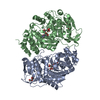
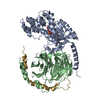
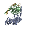
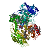
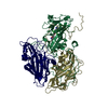
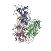
RetinoicacidreceptorRXR-alpha / Retinoid X receptor alpha / Nuclear receptor subfamily 2 group B member 1
Mass: 27114.465 Da / Num. of mol.: 2 / Source method: isolated from a natural source / Source: (natural) Homo sapiens (human) / References: UniProt: P19793
#2: Protein
Nr1h3protein / Putative uncharacterized protein / Nuclear receptor subfamily 1 group H member 3 / Nuclear receptor ...Putative uncharacterized protein / Nuclear receptor subfamily 1 group H member 3 / Nuclear receptor subfamily 1 / group H / member 3 / Nuclear receptor subfamily 1 / group H / member 3 / isoform CRA_a
Mass: 30618.035 Da / Num. of mol.: 2 / Source method: isolated from a natural source / Source: (natural) Mus musculus (house mouse) / References: UniProt: Q91X41, UniProt: Q9Z0Y9*PLUS
In the structure databanks used in Yorodumi, some data are registered as the other names, "COVID-19 virus" and "2019-nCoV". Here are the details of the virus and the list of structure data.
Jan 31, 2019. EMDB accession codes are about to change! (news from PDBe EMDB page)
EMDB accession codes are about to change! (news from PDBe EMDB page)
The allocation of 4 digits for EMDB accession codes will soon come to an end. Whilst these codes will remain in use, new EMDB accession codes will include an additional digit and will expand incrementally as the available range of codes is exhausted. The current 4-digit format prefixed with “EMD-” (i.e. EMD-XXXX) will advance to a 5-digit format (i.e. EMD-XXXXX), and so on. It is currently estimated that the 4-digit codes will be depleted around Spring 2019, at which point the 5-digit format will come into force.
The EM Navigator/Yorodumi systems omit the EMD- prefix.
Related info.:Q: What is EMD? / ID/Accession-code notation in Yorodumi/EM Navigator
Yorodumi is a browser for structure data from EMDB, PDB, SASBDB, etc.
This page is also the successor to EM Navigator detail page, and also detail information page/front-end page for Omokage search.
The word "yorodu" (or yorozu) is an old Japanese word meaning "ten thousand". "mi" (miru) is to see.
Related info.:EMDB / PDB / SASBDB / Comparison of 3 databanks / Yorodumi Search / Aug 31, 2016. New EM Navigator & Yorodumi / Yorodumi Papers / Jmol/JSmol / Function and homology information / Changes in new EM Navigator and Yorodumi
 Movie
Movie Controller


 Open data
 Basic information
 Components
 Keywords
 Function and homology information
 Homo sapiens (human)
 X-RAY DIFFRACTION /
  Authors
 Citation
 Structure visualization
 Downloads & links
 Other downloads


 PDBj
PDBj


 Assembly
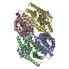
 Mass: 300.435 Da / Num. of mol.: 2 / Source method: obtained synthetically / Formula: C20H28O2
 Mass: 621.088 Da / Num. of mol.: 2 / Source method: obtained synthetically / Formula: C35H32ClF3N2O3
 Mass: 18.015 Da / Num. of mol.: 229 / Source method: isolated from a natural source / Formula: H2O
 Sample preparation
 / Beamline: 17-ID / Wavelength: 1 Å
 Processing