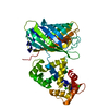
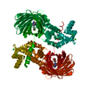
Entry Database : PDB / ID : 3evuTitle Crystal structure of Calcium bound dimeric GCAMP2 Myosin light chain kinase, Green fluorescent protein, Calmodulin-1 chimera Keywords / / / / / Function / homology Function Domain/homology Component
/ / / / / / / / / / / / / / / / / / / / / / / / / / / / / / / / / / / / / / / / / / / / / / / / / / / / / / / / / / / / / / / / / / / / / / / / / / / / / / / / / / / / / / / / / / / / / / / / / / / / / / / / / / / / / / / / / / / / / / / / / / / / / / / / / / / / / / / / / / / / / / / / / / / Biological species Aequorea victoria (jellyfish)Gallus gallus (chicken)Rattus norvegicus (Norway rat)Method / / Resolution : 1.75 Å Authors Wang, Q. / Shui, B. / Kotlikoff, M.I. / Sondermann, H. Journal : Structure / Year : 2008Title : Structural Basis for Calcium Sensing by GCaMP2.Authors : Wang, Q. / Shui, B. / Kotlikoff, M.I. / Sondermann, H. History Deposition Oct 13, 2008 Deposition site / Processing site Revision 1.0 Dec 9, 2008 Provider / Type Revision 1.1 Jul 13, 2011 Group Revision 1.2 Jul 26, 2017 Group Advisory / Data collection ... Advisory / Data collection / Database references / Refinement description / Source and taxonomy / Structure summary Category diffrn_radiation_wavelength / diffrn_source ... diffrn_radiation_wavelength / diffrn_source / entity / entity_name_com / entity_src_gen / pdbx_distant_solvent_atoms / software / struct / struct_ref / struct_ref_seq / struct_ref_seq_dif Item _diffrn_radiation_wavelength.wavelength / _diffrn_source.pdbx_wavelength_list ... _diffrn_radiation_wavelength.wavelength / _diffrn_source.pdbx_wavelength_list / _entity.pdbx_description / _entity.pdbx_ec / _entity.pdbx_fragment / _struct.title / _struct_ref.db_code / _struct_ref.pdbx_align_begin / _struct_ref.pdbx_db_accession / _struct_ref.pdbx_seq_one_letter_code / _struct_ref_seq.db_align_beg / _struct_ref_seq.db_align_end / _struct_ref_seq.pdbx_auth_seq_align_beg / _struct_ref_seq.pdbx_db_accession / _struct_ref_seq.seq_align_beg Revision 1.3 Dec 27, 2023 Group / Database references / Derived calculationsCategory chem_comp_atom / chem_comp_bond ... chem_comp_atom / chem_comp_bond / database_2 / struct_conn / struct_site Item _database_2.pdbx_DOI / _database_2.pdbx_database_accession ... _database_2.pdbx_DOI / _database_2.pdbx_database_accession / _struct_conn.pdbx_leaving_atom_flag / _struct_site.pdbx_auth_asym_id / _struct_site.pdbx_auth_comp_id / _struct_site.pdbx_auth_seq_id Revision 1.4 Nov 20, 2024 Group / Category / pdbx_modification_featureRevision 2.0 Mar 18, 2026 Group / Category / Item
Show all Show less
 Movie
Movie Controller


 Open data
 Basic information
 Components
 Keywords
 Function and homology information
 Aequorea victoria (jellyfish)

 X-RAY DIFFRACTION /
  Authors
 Citation
 Structure visualization
 Downloads & links
 Other downloads
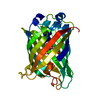


 PDBj
PDBj


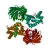
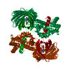
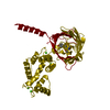
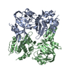
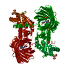
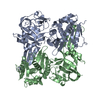
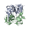
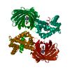
 Assembly
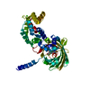


 Mass: 40.078 Da / Num. of mol.: 4 / Source method: obtained synthetically / Formula: Ca
 Mass: 18.015 Da / Num. of mol.: 516 / Source method: isolated from a natural source / Formula: H2O
 Sample preparation
 / Beamline: A1 / Wavelength: 1 Å
 Processing