 Movie
Movie Controller


+ Open data

- Basic information
| Entry | Database: PDB / ID: 3ek7 | |||||||||
|---|---|---|---|---|---|---|---|---|---|---|
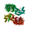
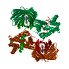
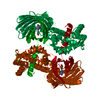
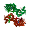
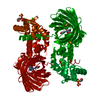
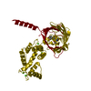
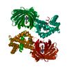
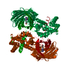
| Title | Calcium-saturated GCaMP2 dimer | |||||||||
 Components Components | Myosin light chain kinase, Green fluorescent protein, Calmodulin chimera | |||||||||
 Keywords Keywords | FLUORESCENT PROTEIN / GECI / GCaMP2 / cpEGFP / calmodulin / M13 peptide / Methylation / Phosphoprotein / SIGNALING PROTEIN | |||||||||
| Function / homology |  Function and homology information Function and homology informationtonic smooth muscle contraction / myosin-light-chain kinase / myosin light chain kinase activity / positive regulation of sarcomere organization / positive regulation of striated muscle contraction / transporter inhibitor activity / : / type 3 metabotropic glutamate receptor binding / establishment of protein localization to membrane / cardiac muscle cell development ...tonic smooth muscle contraction / myosin-light-chain kinase / myosin light chain kinase activity / positive regulation of sarcomere organization / positive regulation of striated muscle contraction / transporter inhibitor activity / : / type 3 metabotropic glutamate receptor binding / establishment of protein localization to membrane / cardiac muscle cell development / response to corticosterone / negative regulation of ryanodine-sensitive calcium-release channel activity / organelle localization by membrane tethering / : / autophagosome membrane docking / regulation of synaptic vesicle exocytosis / negative regulation of calcium ion export across plasma membrane / regulation of ryanodine-sensitive calcium-release channel activity / regulation of cardiac muscle cell action potential / presynaptic endocytosis / cleavage furrow / calcineurin-mediated signaling / nitric-oxide synthase binding / regulation of cell communication by electrical coupling involved in cardiac conduction / adenylate cyclase binding / protein phosphatase activator activity / regulation of synaptic vesicle endocytosis / detection of calcium ion / postsynaptic cytosol / regulation of cardiac muscle contraction / cell surface receptor signaling pathway via JAK-STAT / catalytic complex / heart morphogenesis / phosphatidylinositol 3-kinase binding / calcium channel inhibitor activity / presynaptic cytosol / cellular response to interferon-beta / regulation of release of sequestered calcium ion into cytosol by sarcoplasmic reticulum / stress fiber / titin binding / regulation of cardiac muscle contraction by regulation of the release of sequestered calcium ion / regulation of calcium-mediated signaling / voltage-gated potassium channel complex / calcium channel complex / regulation of heart rate / calyx of Held / nitric-oxide synthase regulator activity / adenylate cyclase activator activity / bioluminescence / protein serine/threonine kinase activator activity / regulation of cytokinesis / spindle microtubule / positive regulation of receptor signaling pathway via JAK-STAT / response to amphetamine / sarcomere / calcium channel regulator activity / generation of precursor metabolites and energy / response to calcium ion / cellular response to type II interferon / G2/M transition of mitotic cell cycle / Schaffer collateral - CA1 synapse / spindle pole / calcium-dependent protein binding / myelin sheath / lamellipodium / synaptic vesicle membrane / growth cone / sperm midpiece / vesicle / transmembrane transporter binding / calmodulin binding / protein domain specific binding / calcium ion binding / centrosome / protein kinase binding / chromatin / protein-containing complex / mitochondrion / nucleoplasm / ATP binding / membrane / metal ion binding / nucleus / cytoplasm / cytosol Similarity search - Function | |||||||||
| Biological species | artificial gene (others)  Aequorea victoria (jellyfish) Aequorea victoria (jellyfish) | |||||||||
| Method |  X-RAY DIFFRACTION / SYNCHROTRON / MOLECULAR REPLACEMENT / molecular replacement / Resolution: 1.85 Å X-RAY DIFFRACTION / SYNCHROTRON / MOLECULAR REPLACEMENT / molecular replacement / Resolution: 1.85 Å | |||||||||
 Authors Authors | Akerboom, J. / Velez Rivera, J.D. / Looger, L.L. / Schreiter, E.R. | |||||||||
 Citation Citation | Journal: J.Biol.Chem. / Year: 2009 Title: Crystal Structures of the GCaMP Calcium Sensor Reveal the Mechanism of Fluorescence Signal Change and Aid Rational Design Authors: Akerboom, J. / Rivera, J.D. / Guilbe, M.M. / Malave, E.C. / Hernandez, H.H. / Tian, L. / Hires, S.A. / Marvin, J.S. / Looger, L.L. / Schreiter, E.R. #1: Journal: Acta Crystallogr.,Sect.F / Year: 2008Title: Crystallization and preliminary X-ray characterization of the genetically encoded fluorescent calcium indicator protein GCaMP2 Authors: M Rodriguez Guilbe, M. / Alfaro Malave, E.C. / Akerboom, J. / Marvin, J.S. / Looger, L.L. / Schreiter, E.R. | |||||||||
| History |
|
- Structure visualization
| Structure viewer | Molecule: MolmilJmol/JSmol |
|---|
- Downloads & links
-Download
| PDBx/mmCIF format | 3ek7.cif.gz | 100.7 KB | Display | PDBx/mmCIF format |
|---|---|---|---|---|
| PDB format | pdb3ek7.ent.gz | 74.3 KB | Display | PDB format |
| PDBx/mmJSON format | 3ek7.json.gz | Tree view | PDBx/mmJSON format | |
| Others |  Other downloads Other downloads |
-Validation report
| Arichive directory | https://data.pdbj.org/pub/pdb/validation_reports/ek/3ek7ftp://data.pdbj.org/pub/pdb/validation_reports/ek/3ek7 | HTTPS FTP |
|---|
-Related structure data
| Related structure data | 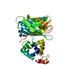 3ek4C 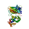 3ek8C 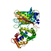 3ekhC 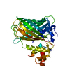 3ekjC  1cdlS  1emaS C: citing same article ( S: Starting model for refinement |
|---|---|
| Similar structure data |


-Links
 PDBj
PDBj


- Assembly
| Deposited unit | 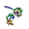
| ||||||||
|---|---|---|---|---|---|---|---|---|---|
| 1 | 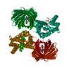
| ||||||||
| Unit cell |
|
-Components
| #1: Protein | Mass: 50704.586 Da / Num. of mol.: 1 Source method: isolated from a genetically manipulated source Details: calcium-sensor; protein contains circular permuted EGFP, the M13 fragment of myosin light chain kinase, rat calmodulin and a 6x His-tag from the pRSETA vector Source: (gene. exp.) artificial gene (others), (gene. exp.) Aequorea victoria (jellyfish), (gene. exp.) Plasmid: pRSETA / Gene: GFP, Calm1, Calm, Cam, Cam1, CaMI / Production host:  References: UniProt: P42212, UniProt: P0DP29, UniProt: P11799*PLUS | ||||||
|---|---|---|---|---|---|---|---|
| #2: Chemical | ChemComp-CA /   Mass: 40.078 Da / Num. of mol.: 4 / Source method: obtained synthetically / Formula: Ca Mass: 40.078 Da / Num. of mol.: 4 / Source method: obtained synthetically / Formula: Ca#3: Water | ChemComp-HOH / |  Mass: 18.015 Da / Num. of mol.: 198 / Source method: isolated from a natural source / Formula: H2O Mass: 18.015 Da / Num. of mol.: 198 / Source method: isolated from a natural source / Formula: H2OHas protein modification | Y | Sequence details | RESIDUE SER65 HAS BEEN MUTATED TO GLY. RESIDUES GLY65, TYR66, AND GLY67 CONSTITUTE THE CHROMOPHORE ...RESIDUE SER65 HAS BEEN MUTATED TO GLY. RESIDUES GLY65, TYR66, AND GLY67 CONSTITUTE | |
-Experimental details
-Experiment
| Experiment | Method: X-RAY DIFFRACTION / Number of used crystals: 1 |
|---|
- Sample preparation
| Crystal | Density Matthews: 1.99 Å3/Da / Density % sol: 38.06 % |
|---|---|
| Crystal grow | Temperature: 298 K / Method: vapor diffusion, hanging drop / pH: 8.5 Details: 0.2 M lithium sulfate monohydrate, 0.1 M Tris-HCl pH 8.5, 30%(w/v) polyethylene glycol 4000, VAPOR DIFFUSION, HANGING DROP, temperature 298K |
-Data collection
| Diffraction | Mean temperature: 100 K |
|---|---|
| Diffraction source | Source: SYNCHROTRON / Site: APS  / Beamline: 31-ID / Wavelength: 0.9793 Å / Beamline: 31-ID / Wavelength: 0.9793 Å |
| Detector | Type: MAR CCD 165 mm / Detector: CCD / Date: Jul 28, 2008 |
| Radiation | Monochromator: Diamond(1,1,1) / Protocol: SINGLE WAVELENGTH / Monochromatic (M) / Laue (L): M / Scattering type: x-ray |
| Radiation wavelength | Wavelength: 0.9793 Å / Relative weight: 1 |
| Reflection | Resolution: 1.85→67.73 Å / Num. obs: 36788 / % possible obs: 98.49 % / Observed criterion σ(F): 0 / Observed criterion σ(I): 0 / Redundancy: 7.5 % / Rsym value: 0.076 |
| Reflection shell | Resolution: 1.85→1.9 Å / Redundancy: 7.6 % / Mean I/σ(I) obs: 5.1 / Rsym value: 0.42 / % possible all: 97.6 |
-Phasing
| Phasing | Method: molecular replacement |
|---|
- Processing
| Software |
| ||||||||||||||||||||||||||||||||||||||||||||||||||||||||||||||||||||||||||||||||||||||||||||||||||||||||||||||||||||||||||||||||||||||||||||||||||||||||||||||||||||||||||
|---|---|---|---|---|---|---|---|---|---|---|---|---|---|---|---|---|---|---|---|---|---|---|---|---|---|---|---|---|---|---|---|---|---|---|---|---|---|---|---|---|---|---|---|---|---|---|---|---|---|---|---|---|---|---|---|---|---|---|---|---|---|---|---|---|---|---|---|---|---|---|---|---|---|---|---|---|---|---|---|---|---|---|---|---|---|---|---|---|---|---|---|---|---|---|---|---|---|---|---|---|---|---|---|---|---|---|---|---|---|---|---|---|---|---|---|---|---|---|---|---|---|---|---|---|---|---|---|---|---|---|---|---|---|---|---|---|---|---|---|---|---|---|---|---|---|---|---|---|---|---|---|---|---|---|---|---|---|---|---|---|---|---|---|---|---|---|---|---|---|---|---|
| Refinement | Method to determine structure: MOLECULAR REPLACEMENT Starting model: PDB entries 1EMA and 1CDL Resolution: 1.85→20.67 Å / Cor.coef. Fo:Fc: 0.951 / Cor.coef. Fo:Fc free: 0.923 / Occupancy max: 1 / Occupancy min: 0.4 / SU B: 3.064 / SU ML: 0.095 / Cross valid method: THROUGHOUT / ESU R: 0.15 / ESU R Free: 0.146 / Stereochemistry target values: MAXIMUM LIKELIHOOD / Details: HYDROGENS HAVE BEEN ADDED IN THE RIDING POSITIONS
| ||||||||||||||||||||||||||||||||||||||||||||||||||||||||||||||||||||||||||||||||||||||||||||||||||||||||||||||||||||||||||||||||||||||||||||||||||||||||||||||||||||||||||
| Solvent computation | Ion probe radii: 0.8 Å / Shrinkage radii: 0.8 Å / VDW probe radii: 1.2 Å / Solvent model: MASK | ||||||||||||||||||||||||||||||||||||||||||||||||||||||||||||||||||||||||||||||||||||||||||||||||||||||||||||||||||||||||||||||||||||||||||||||||||||||||||||||||||||||||||
| Displacement parameters | Biso mean: 20.837 Å2
| ||||||||||||||||||||||||||||||||||||||||||||||||||||||||||||||||||||||||||||||||||||||||||||||||||||||||||||||||||||||||||||||||||||||||||||||||||||||||||||||||||||||||||
| Refinement step | Cycle: LAST / Resolution: 1.85→20.67 Å
| ||||||||||||||||||||||||||||||||||||||||||||||||||||||||||||||||||||||||||||||||||||||||||||||||||||||||||||||||||||||||||||||||||||||||||||||||||||||||||||||||||||||||||
| Refine LS restraints |
| ||||||||||||||||||||||||||||||||||||||||||||||||||||||||||||||||||||||||||||||||||||||||||||||||||||||||||||||||||||||||||||||||||||||||||||||||||||||||||||||||||||||||||
| LS refinement shell | Resolution: 1.85→1.898 Å / Total num. of bins used: 20
|
