 Movie
Movie Controller


+ Open data

- Basic information
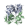
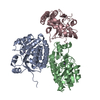
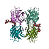
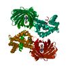
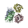
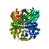
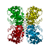
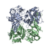
| Entry | Database: PDB / ID: 1ef0 | ||||||
|---|---|---|---|---|---|---|---|
| Title | CRYSTAL STRUCTURE OF PI-SCEI MINIPRECURSOR | ||||||
 Components Components | PI-SCEI ENDONUCLEASE | ||||||
 Keywords Keywords | HYDROLASE / endonuclease / protein splicing / mini-precursor | ||||||
| Function / homology |  Function and homology information Function and homology informationInsulin receptor recycling / Transferrin endocytosis and recycling / ROS and RNS production in phagocytes / Golgi lumen acidification / Amino acids regulate mTORC1 / vacuolar proton-transporting V-type ATPase, V1 domain / endosomal lumen acidification / intron homing / intein-mediated protein splicing / vacuolar acidification ...Insulin receptor recycling / Transferrin endocytosis and recycling / ROS and RNS production in phagocytes / Golgi lumen acidification / Amino acids regulate mTORC1 / vacuolar proton-transporting V-type ATPase, V1 domain / endosomal lumen acidification / intron homing / intein-mediated protein splicing / vacuolar acidification / fungal-type vacuole membrane / proton-transporting ATPase activity, rotational mechanism / H+-transporting two-sector ATPase / ATP metabolic process / proton transmembrane transport / endonuclease activity / Hydrolases; Acting on ester bonds / Golgi membrane / hydrolase activity / mRNA binding / DNA binding / ATP binding Similarity search - Function | ||||||
| Biological species |  | ||||||
| Method |  X-RAY DIFFRACTION / SYNCHROTRON / Resolution: 2.1 Å X-RAY DIFFRACTION / SYNCHROTRON / Resolution: 2.1 Å | ||||||
 Authors Authors | Poland, B.W. / Xu, M.-Q. / Quiocho, F.A. | ||||||
 Citation Citation | Journal: J.Biol.Chem. / Year: 2000 Title: Structural insights into the protein splicing mechanism of PI-SceI. Authors: Poland, B.W. / Xu, M.Q. / Quiocho, F.A. #1: Journal: Cell(Cambridge,Mass.) / Year: 1997Title: Crystal Structure of PI-SceI, a Homing Endonuclease with Protein Splicing Activity Authors: Duan, X. / Gimble, F.S. / Quiocho, F.A. #2: Journal: Science / Year: 1990Title: Protein Splicing Converts the Yeast TFP1 Gene Product to the 69-kD Subunit of the Vacuolar H+-Adenosine Triphosphatase Authors: Kane, P.M. / Yamashiro, C.T. / Wolczyk, D.F. / Neff, N. / Goebl, M. / Stevens, T.H. #3: Journal: Chem.Soc.Rev. / Year: 1998Title: The chemical basis of protein splicing. Authors: Paulus, H. | ||||||
| History |
|
- Structure visualization
| Structure viewer | Molecule: MolmilJmol/JSmol |
|---|
- Downloads & links
-Download
| PDBx/mmCIF format | 1ef0.cif.gz | 179.6 KB | Display | PDBx/mmCIF format |
|---|---|---|---|---|
| PDB format | pdb1ef0.ent.gz | 142.2 KB | Display | PDB format |
| PDBx/mmJSON format | 1ef0.json.gz | Tree view | PDBx/mmJSON format | |
| Others |  Other downloads Other downloads |
-Validation report
| Arichive directory | https://data.pdbj.org/pub/pdb/validation_reports/ef/1ef0ftp://data.pdbj.org/pub/pdb/validation_reports/ef/1ef0 | HTTPS FTP |
|---|
-Related structure data
| Similar structure data |
|---|


-Links
 PDBj
PDBj


- Assembly
| Deposited unit | 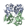
| ||||||||
|---|---|---|---|---|---|---|---|---|---|
| 1 |
| ||||||||
| 2 | 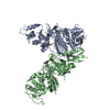
| ||||||||
| Unit cell |
|
-Components
| #1: Protein | Mass: 51803.812 Da / Num. of mol.: 2 Source method: isolated from a genetically manipulated source Details: FROM THE VACUOLAR ATPASE SUBUNIT Source: (gene. exp.) Plasmid: PVMA29 / Production host:  References: GenBank: 172907, UniProt: P17255*PLUS, EC: 3.6.1.34 #2: Chemical |   Mass: 65.409 Da / Num. of mol.: 2 / Source method: obtained synthetically / Formula: Zn Mass: 65.409 Da / Num. of mol.: 2 / Source method: obtained synthetically / Formula: Zn#3: Water | ChemComp-HOH / |  Mass: 18.015 Da / Num. of mol.: 408 / Source method: isolated from a natural source / Formula: H2O Mass: 18.015 Da / Num. of mol.: 408 / Source method: isolated from a natural source / Formula: H2O |
|---|
-Experimental details
-Experiment
| Experiment | Method: X-RAY DIFFRACTION / Number of used crystals: 1 |
|---|
- Sample preparation
| Crystal | Density Matthews: 2.51 Å3/Da / Density % sol: 50.93 % | ||||||||||||||||||||||||||||||||||||||||||
|---|---|---|---|---|---|---|---|---|---|---|---|---|---|---|---|---|---|---|---|---|---|---|---|---|---|---|---|---|---|---|---|---|---|---|---|---|---|---|---|---|---|---|---|
| Crystal grow | Temperature: 298 K / Method: vapor diffusion, hanging drop / pH: 8.5 Details: PEG 3350, Cadmium chloride, magnesium chloride, mercaptoethanol, tric-HCl, pH 8.5, VAPOR DIFFUSION, HANGING DROP, temperature 298K | ||||||||||||||||||||||||||||||||||||||||||
| Crystal grow | *PLUS Details: drop consists of equal volume of protein and reservoir solutions | ||||||||||||||||||||||||||||||||||||||||||
| Components of the solutions | *PLUS
|
-Data collection
| Diffraction | Mean temperature: 100 K |
|---|---|
| Diffraction source | Source: SYNCHROTRON / Site: NSLS  / Beamline: X4A / Wavelength: 0.9791 / Beamline: X4A / Wavelength: 0.9791 |
| Detector | Type: RIGAKU RAXIS / Detector: IMAGE PLATE / Date: Oct 1, 1998 |
| Radiation | Protocol: SINGLE WAVELENGTH / Monochromatic (M) / Laue (L): M / Scattering type: x-ray |
| Radiation wavelength | Wavelength: 0.9791 Å / Relative weight: 1 |
| Reflection | Resolution: 2.1→20 Å / Num. all: 61089 / Num. obs: 56914 / % possible obs: 93.3 % / Observed criterion σ(F): 0 / Observed criterion σ(I): 2 / Redundancy: 6.8 % / Rmerge(I) obs: 0.101 / Net I/σ(I): 8.6 |
| Reflection shell | Resolution: 2.1→20 Å / Redundancy: 6.8 % / Rmerge(I) obs: 0.101 / Num. unique all: 61089 / % possible all: 93.3 |
| Reflection | *PLUS Num. obs: 61089 / Num. measured all: 419279 |
| Reflection shell | *PLUS % possible obs: 93.3 % |
- Processing
| Software |
| |||||||||||||||||||||||||
|---|---|---|---|---|---|---|---|---|---|---|---|---|---|---|---|---|---|---|---|---|---|---|---|---|---|---|
| Refinement | Resolution: 2.1→20 Å / σ(F): 0 / σ(I): 0 / Stereochemistry target values: Engh & Huber
| |||||||||||||||||||||||||
| Refinement step | Cycle: LAST / Resolution: 2.1→20 Å
| |||||||||||||||||||||||||
| Refine LS restraints |
| |||||||||||||||||||||||||
| Software | *PLUS Name: 'CNS' / Classification: refinement | |||||||||||||||||||||||||
| Refine LS restraints | *PLUS
|
