 Movie
Movie Controller

[English] 日本語
 Yorodumi
- PDB-3euk: Crystal structure of MukE-MukF(residues 292-443)-MukB(head domain... -
+ Open data

- Basic information
| Entry | Database: PDB / ID: 3euk | ||||||
|---|---|---|---|---|---|---|---|
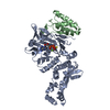
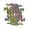
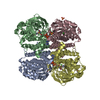
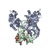
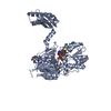
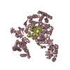
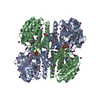
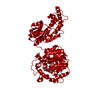
| Title | Crystal structure of MukE-MukF(residues 292-443)-MukB(head domain)-ATPgammaS complex, asymmetric dimer | ||||||
 Components Components |
| ||||||
 Keywords Keywords | CELL CYCLE / MukB / MukE / MukF / Chromosome condensation / condensin / SMC / non-SMC subunit / ABC-type ATPase / WHD / ATP-binding / Cell division / Chromosome partition / DNA condensation / DNA-binding / Nucleotide-binding | ||||||
| Function / homology |  Function and homology information Function and homology informationnucleoid / chromosome condensation / chromosome segregation / DNA replication / cell division / calcium ion binding / DNA binding / ATP binding / cytoplasm Similarity search - Function | ||||||
| Biological species |  Haemophilus ducreyi (bacteria) Haemophilus ducreyi (bacteria) | ||||||
| Method |  X-RAY DIFFRACTION / SYNCHROTRON / Resolution: 4 Å X-RAY DIFFRACTION / SYNCHROTRON / Resolution: 4 Å | ||||||
 Authors Authors | Woo, J.S. / Lim, J.H. / Shin, H.C. / Oh, B.H. | ||||||
 Citation Citation | Journal: Cell(Cambridge,Mass.) / Year: 2009 Title: Structural studies of a bacterial condensin complex reveal ATP-dependent disruption of intersubunit interactions. Authors: Woo, J.S. / Lim, J.H. / Shin, H.C. / Suh, M.K. / Ku, B. / Lee, K.H. / Joo, K. / Robinson, H. / Lee, J. / Park, S.Y. / Ha, N.C. / Oh, B.H. | ||||||
| History |
|
- Structure visualization
| Structure viewer | Molecule: MolmilJmol/JSmol |
|---|
- Downloads & links
-Download
| PDBx/mmCIF format | 3euk.cif.gz | 466.6 KB | Display | PDBx/mmCIF format |
|---|---|---|---|---|
| PDB format | pdb3euk.ent.gz | 374.7 KB | Display | PDB format |
| PDBx/mmJSON format | 3euk.json.gz | Tree view | PDBx/mmJSON format | |
| Others |  Other downloads Other downloads |
-Validation report
| Summary document | 3euk_validation.pdf.gz | 1.2 MB | Display | wwPDB validaton report |
|---|---|---|---|---|
| Full document | 3euk_full_validation.pdf.gz | 1.4 MB | Display | |
| Data in XML | 3euk_validation.xml.gz | 102.3 KB | Display | |
| Data in CIF | 3euk_validation.cif.gz | 134.6 KB | Display | |
| Arichive directory | https://data.pdbj.org/pub/pdb/validation_reports/eu/3eukftp://data.pdbj.org/pub/pdb/validation_reports/eu/3euk | HTTPS FTP |
-Related structure data


-Links
 PDBj
PDBj
- Assembly
| Deposited unit | 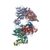
| ||||||||
|---|---|---|---|---|---|---|---|---|---|
| 1 | 
| ||||||||
| 2 | 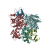
| ||||||||
| Unit cell |
|
-Components
| #1: Protein | Mass: 54102.809 Da / Num. of mol.: 4 / Fragment: Head domain / Mutation: E1435Q Source method: isolated from a genetically manipulated source Source: (gene. exp.) Haemophilus ducreyi (strain 35000HP / ATCC 700724) (bacteria)Gene: mukB, HD_1582 / Plasmid: pPROEX / Production host: #2: Protein | Mass: 17334.508 Da / Num. of mol.: 2 / Fragment: residues 292-443 Source method: isolated from a genetically manipulated source Source: (gene. exp.) Haemophilus ducreyi (bacteria) / Gene: mukF, HD_1585 / Plasmid: pET30a / Production host: #3: Protein | Mass: 26815.508 Da / Num. of mol.: 2 Source method: isolated from a genetically manipulated source Source: (gene. exp.) Haemophilus ducreyi (bacteria) / Gene: mukE, HD_1584 / Plasmid: pET30a / Production host: #4: Chemical | ChemComp-AGS /   Mass: 523.247 Da / Num. of mol.: 4 / Source method: obtained synthetically / Formula: C10H16N5O12P3S / Comment: ATP-gamma-S, energy-carrying molecule analogue*YM Mass: 523.247 Da / Num. of mol.: 4 / Source method: obtained synthetically / Formula: C10H16N5O12P3S / Comment: ATP-gamma-S, energy-carrying molecule analogue*YM#5: Chemical | ChemComp-MG /   Mass: 24.305 Da / Num. of mol.: 4 / Source method: obtained synthetically / Formula: Mg Mass: 24.305 Da / Num. of mol.: 4 / Source method: obtained synthetically / Formula: MgSequence details | THESE RESIDUES ARE NATURAL GENETIC VARIANTS, POLYMORPHI | |
|---|
-Experimental details
-Experiment
| Experiment | Method: X-RAY DIFFRACTION / Number of used crystals: 1 |
|---|
- Sample preparation
| Crystal | Density Matthews: 3.46 Å3/Da / Density % sol: 64.46 % / Mosaicity: 0.412 ° |
|---|---|
| Crystal grow | Temperature: 296 K / Method: hanging drop / pH: 8 Details: PEG 10000, MgCl2, pH 8.0, hanging drop, temperature 296K |
-Data collection
| Diffraction | Mean temperature: 100 K | |||||||||||||||||||||||||||||||||||||||||||||||||||||||||||||||||||||||||||||
|---|---|---|---|---|---|---|---|---|---|---|---|---|---|---|---|---|---|---|---|---|---|---|---|---|---|---|---|---|---|---|---|---|---|---|---|---|---|---|---|---|---|---|---|---|---|---|---|---|---|---|---|---|---|---|---|---|---|---|---|---|---|---|---|---|---|---|---|---|---|---|---|---|---|---|---|---|---|---|
| Diffraction source | Source: SYNCHROTRON / Site: Photon Factory  / Beamline: BL-5A / Wavelength: 1 Å / Beamline: BL-5A / Wavelength: 1 Å | |||||||||||||||||||||||||||||||||||||||||||||||||||||||||||||||||||||||||||||
| Detector | Type: ADSC QUANTUM 210r / Detector: CCD / Date: Mar 9, 2007 | |||||||||||||||||||||||||||||||||||||||||||||||||||||||||||||||||||||||||||||
| Radiation | Protocol: SINGLE WAVELENGTH / Monochromatic (M) / Laue (L): M / Scattering type: x-ray | |||||||||||||||||||||||||||||||||||||||||||||||||||||||||||||||||||||||||||||
| Radiation wavelength | Wavelength: 1 Å / Relative weight: 1 | |||||||||||||||||||||||||||||||||||||||||||||||||||||||||||||||||||||||||||||
| Reflection | Resolution: 4→30 Å / Num. obs: 34959 / % possible obs: 92.8 % / Redundancy: 6 % / Rmerge(I) obs: 0.077 / Χ2: 0.84 / Net I/σ(I): 11.576 | |||||||||||||||||||||||||||||||||||||||||||||||||||||||||||||||||||||||||||||
| Reflection shell |
|
- Processing
| Software |
| ||||||||||||||||||||
|---|---|---|---|---|---|---|---|---|---|---|---|---|---|---|---|---|---|---|---|---|---|
| Refinement | Resolution: 4→20 Å / Occupancy max: 1 / Occupancy min: 1 / FOM work R set: 0.756 / σ(F): 0
| ||||||||||||||||||||
| Solvent computation | Bsol: 35.472 Å2 | ||||||||||||||||||||
| Displacement parameters | Biso max: 200 Å2 / Biso mean: 130.265 Å2 / Biso min: 26.49 Å2
| ||||||||||||||||||||
| Refinement step | Cycle: LAST / Resolution: 4→20 Å
| ||||||||||||||||||||
| Refine LS restraints |
| ||||||||||||||||||||
| Xplor file |
|
