- PDB-3etn: Crystal structure of putative phosphosugar isomerase involved in ... -
+
Open data
ID or keywords:
Loading...
-
Basic information
Entry
Database: PDB / ID: 3etn
Title
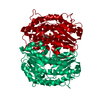
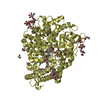
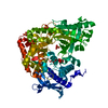
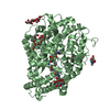
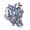
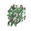
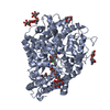
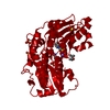
Crystal structure of putative phosphosugar isomerase involved in capsule formation (YP_209877.1) from Bacteroides fragilis NCTC 9343 at 1.70 A resolution
Components
putative phosphosugar isomerase involved in capsule formation
Keywords
ISOMERASE / YP_209877.1 / putative phosphosugar isomerase involved in capsule formation / Structural Genomics / Joint Center for Structural Genomics / JCSG / Protein Structure Initiative / PSI-2 / Unknown function
Function / homology
Function and homology information
carbohydrate derivative metabolic process / carbohydrate derivative binding / isomerase activity Similarity search - Function
Type: MARMOSAIC 325 mm CCD / Detector: CCD / Date: Aug 1, 2008 / Details: Flat mirror (vertical focusing)
Radiation
Monochromator: Single crystal Si(111) bent monochromator (horizontal focusing) Protocol: MAD / Monochromatic (M) / Laue (L): M / Scattering type: x-ray
Radiation wavelength
ID
Wavelength (Å)
Relative weight
1
0.91837
1
2
0.9791
1
3
0.97849
1
Reflection
Resolution: 1.7→29.223 Å / Num. obs: 88793 / % possible obs: 99.8 % / Redundancy: 4.5 % / Biso Wilson estimate: 19.097 Å2 / Rmerge(I) obs: 0.103 / Rsym value: 0.103 / Net I/σ(I): 5.554
Reflection shell
Diffraction-ID: 1
Resolution (Å)
Redundancy (%)
Rmerge(I) obs
Mean I/σ(I) obs
Num. measured all
Num. unique all
Rsym value
% possible all
1.7-1.74
4.5
0.819
0.9
29023
6471
0.819
100
1.74-1.79
4.5
0.662
1.2
28629
6371
0.662
100
1.79-1.84
4.5
0.553
1.4
27797
6169
0.553
100
1.84-1.9
4.5
0.438
1.8
27037
5982
0.438
100
1.9-1.96
4.5
0.371
1.9
25876
5798
0.371
100
1.96-2.03
4.5
0.289
2.6
25441
5615
0.289
100
2.03-2.11
4.5
0.231
3.2
24716
5457
0.231
100
2.11-2.19
4.5
0.181
4.1
23751
5243
0.181
100
2.19-2.29
4.3
0.169
3.6
20881
4881
0.169
97.2
2.29-2.4
4.5
0.133
5.4
21772
4807
0.133
100
2.4-2.53
4.5
0.117
5.8
20948
4620
0.117
100
2.53-2.69
4.5
0.105
6.3
19631
4328
0.105
100
2.69-2.87
4.5
0.094
6.9
18628
4118
0.094
100
2.87-3.1
4.5
0.084
7.4
17343
3826
0.084
100
3.1-3.4
4.5
0.067
9.3
15989
3550
0.067
100
3.4-3.8
4.5
0.058
10.7
14390
3218
0.058
100
3.8-4.39
4.5
0.052
10.6
12747
2851
0.052
100
4.39-5.38
4.4
0.047
11.9
10794
2447
0.047
100
5.38-7.6
4.3
0.046
13.9
8294
1938
0.046
100
7.6-29.223
4
0.039
15
4398
1103
0.039
98.1
-
Phasing
Phasing
Method: MAD
-
Processing
Software
Name
Version
Classification
NB
REFMAC
5.5.0053
refinement
PHENIX
refinement
SHELX
phasing
MolProbity
3beta29
modelbuilding
SCALA
3.2.5
datascaling
PDB_EXTRACT
3.006
dataextraction
MOSFLM
datareduction
SHELXD
phasing
autoSHARP
phasing
Refinement
Method to determine structure: MAD / Resolution: 1.7→29.223 Å / Cor.coef. Fo:Fc: 0.958 / Cor.coef. Fo:Fc free: 0.949 / Occupancy max: 1 / Occupancy min: 0.25 / SU B: 4.785 / SU ML: 0.068 / TLS residual ADP flag: LIKELY RESIDUAL / Cross valid method: THROUGHOUT / σ(F): 0 / ESU R: 0.106 / ESU R Free: 0.099 Stereochemistry target values: MAXIMUM LIKELIHOOD WITH PHASES Details: 1. HYDROGENS HAVE BEEN ADDED IN THE RIDING POSITIONS. 2. A MET-INHIBITION PROTOCOL WAS USED FOR SELENOMETHIONINE INCORPORATION DURING PROTEIN EXPRESSION. THE OCCUPANCY OF THE SE ATOMS IN THE ...Details: 1. HYDROGENS HAVE BEEN ADDED IN THE RIDING POSITIONS. 2. A MET-INHIBITION PROTOCOL WAS USED FOR SELENOMETHIONINE INCORPORATION DURING PROTEIN EXPRESSION. THE OCCUPANCY OF THE SE ATOMS IN THE MSE RESIDUES WAS REDUCED TO 0.75 FOR THE REDUCED SCATTERING POWER DUE TO PARTIAL S-MET INCORPORATION. 3. ATOM RECORDS CONTAIN RESIDUAL B FACTORS ONLY. 4. CMK MOLECULES (CMP-2-keto-3-deoxy-octulosonic acid)
Rfactor
Num. reflection
% reflection
Selection details
Rfree
0.197
4437
5 %
RANDOM
Rwork
0.172
-
-
-
obs
0.173
88524
99.56 %
-
Solvent computation
Ion probe radii: 0.8 Å / Shrinkage radii: 0.8 Å / VDW probe radii: 1.2 Å / Solvent model: MASK
In the structure databanks used in Yorodumi, some data are registered as the other names, "COVID-19 virus" and "2019-nCoV". Here are the details of the virus and the list of structure data.
Jan 31, 2019. EMDB accession codes are about to change! (news from PDBe EMDB page)
EMDB accession codes are about to change! (news from PDBe EMDB page)
The allocation of 4 digits for EMDB accession codes will soon come to an end. Whilst these codes will remain in use, new EMDB accession codes will include an additional digit and will expand incrementally as the available range of codes is exhausted. The current 4-digit format prefixed with “EMD-” (i.e. EMD-XXXX) will advance to a 5-digit format (i.e. EMD-XXXXX), and so on. It is currently estimated that the 4-digit codes will be depleted around Spring 2019, at which point the 5-digit format will come into force.
The EM Navigator/Yorodumi systems omit the EMD- prefix.
Related info.:Q: What is EMD? / ID/Accession-code notation in Yorodumi/EM Navigator
Yorodumi is a browser for structure data from EMDB, PDB, SASBDB, etc.
This page is also the successor to EM Navigator detail page, and also detail information page/front-end page for Omokage search.
The word "yorodu" (or yorozu) is an old Japanese word meaning "ten thousand". "mi" (miru) is to see.
Related info.:EMDB / PDB / SASBDB / Comparison of 3 databanks / Yorodumi Search / Aug 31, 2016. New EM Navigator & Yorodumi / Yorodumi Papers / Jmol/JSmol / Function and homology information / Changes in new EM Navigator and Yorodumi
 Movie
Movie Controller

 Yorodumi
 Open data
 Basic information
 Components
 Keywords
 Function and homology information
 Bacteroides fragilis NCTC 9343 (bacteria)
 X-RAY DIFFRACTION /
  Authors
 Citation
 Structure visualization
 Downloads & links
 Other downloads
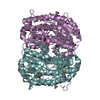

 PDBj
PDBj Assembly
 Mass: 543.373 Da / Num. of mol.: 4 / Source method: obtained synthetically / Formula: C17H26N3O15P
 Mass: 62.068 Da / Num. of mol.: 9 / Source method: obtained synthetically / Formula: C2H6O2
 Mass: 18.015 Da / Num. of mol.: 725 / Source method: isolated from a natural source / Formula: H2O
 Sample preparation
 / Beamline: BL11-1 / Wavelength: 0.91837,0.97910,0.97849
 Processing